
|
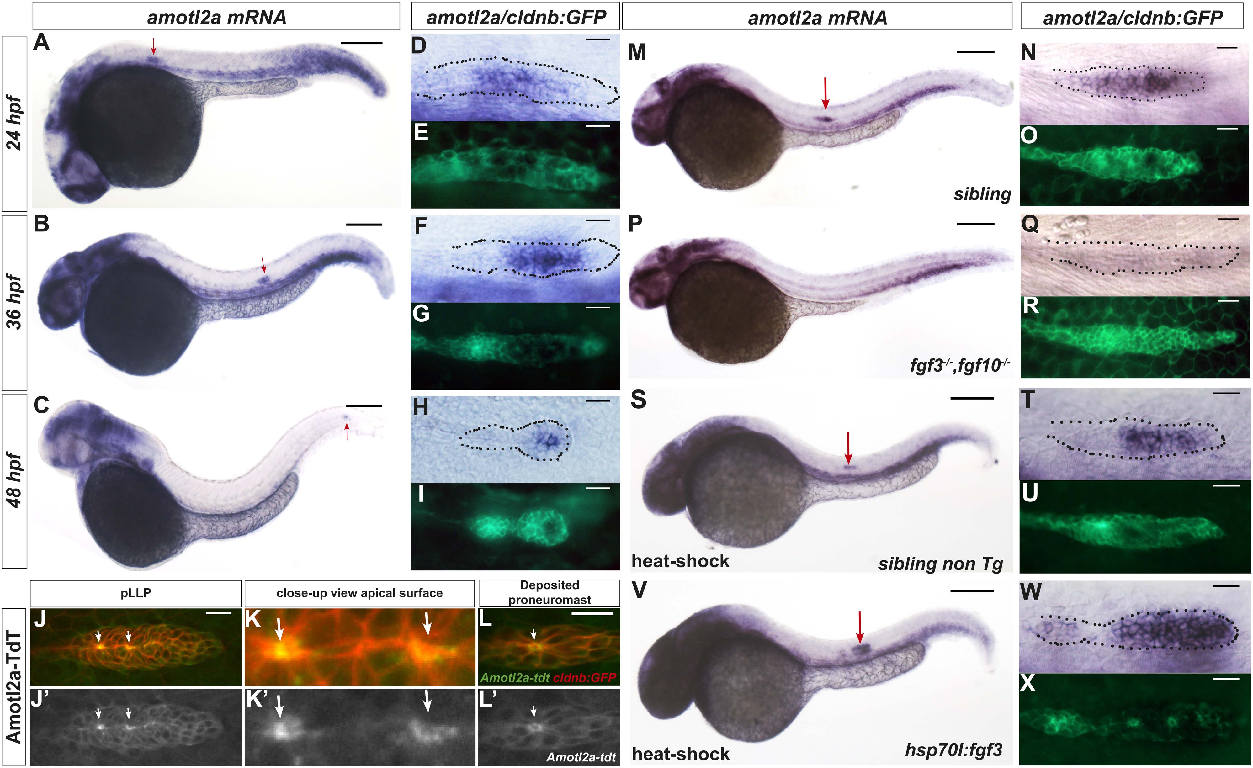
Fig. 1 amotl2a is expressed in the pLLP and localizes at the cell apical side.
(A?I) cldnb:gfp embryos stained with an amotl2a antisense RNA probe and an anti-GFP antibody (E, G, I) at the indicated stages. Red arrows indicate the posterior lateral line primordium (pLLP). (D?I) Close-up views of the pLLP. (J?L′) Maximum intensity projection (MIP) of Z-stacks of the pLLP (J?J′) and a recently deposited neuromast (L?L′) in cldnb:gfp embryos injected with amotl2a-TdT mRNA. (K?K′) Close-up views of (J?J′). White arrows indicate rosette centers. Colors have been inverted. (M?X) 30 hpf cldnb:gfp embryos stained with an amotl2a in situ hybridization (ISH) probe and an anti-GFP antibody (O, R, U, X) in the indicated genetic background. The right column shows the primordium at higher magnification. In all figures, scale bars correspond to 200 �m for whole-embryo images and 20 �m in close up views of the pLLP. In all figures, n is the total number of embryos/primordia analysed and N is the number of biological replicates (Figure 1?figure supplement 1, Figure 1?source data 1).