
|
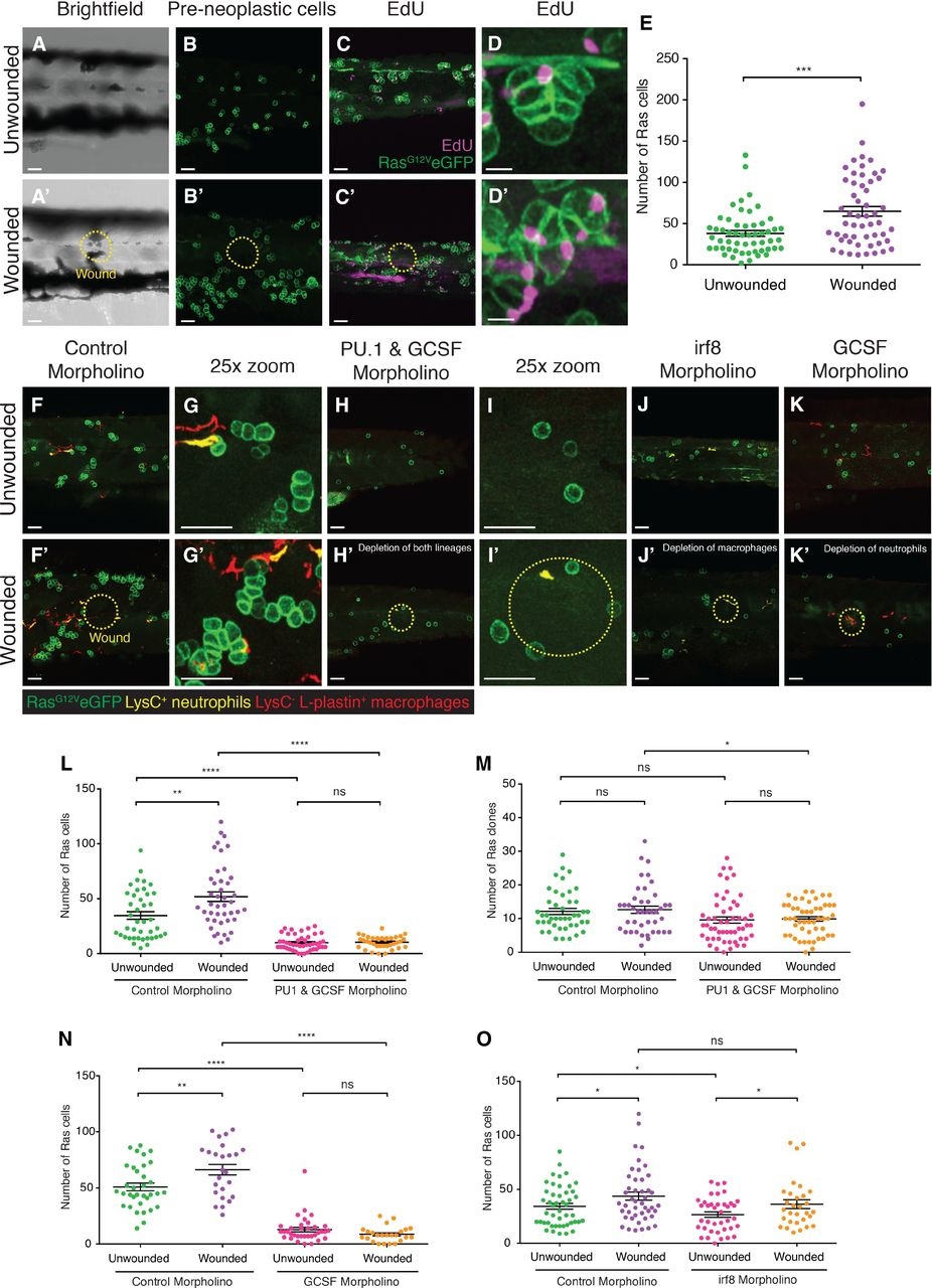
Fig. 3
Wounding leads to increased proliferation of pre-neoplastic cells.
A–D′ RasG12VeGFP larvae were left unwounded (A–D) or laserwounded (yellow dotted line) at 2 dpf just dorsal to the cloaca (A′–D′). Larvae were left to grow for 3 days before being fixed and analysed for preneoplastic cell number (n = 30 larvae in each group). EdU accumulation (purple) is shown at low magnification (C and C′) and a representative preneoplastic cell clone is shown (D and D′).
E. Graph illustrating preneoplastic cell numbers in unwounded versus wounded larvae (n = 27 of each group). Data from three independent experiments showed the same level of significance. ***P d 0.001.
F–G′ Images of larvae injected with control morpholino at the onecell stage, and either left unwounded (F, high magnification: G) or laserwounded at 3 dpf (F′, high magnification: G′) and fixed at 5 dpf.
H–J′ Larvae injected with a combination of PU1 and GCSF morpholinos at the onecell stage, left unwounded (H, high magnification: I) or laserwounded at 3 dpf (H′, high magnification: I′) and fixed at 5 dpf. Images of larvae injected with irf8 morpholino, and either unwounded (J) or laserwounded at 3 dpf (J′) before subsequent fixation at 5 dpf.
K, K′ Larvae injected with GCSF morpholino, and either left unwounded (K) or laserwounded at 3 dpf (K′) and fixed at 5 dpf.
L, M Graphs to show the total number of Ras+ cells and clones (respectively) at 5 dpf after PU1 and GCSF morpholinos.
N, O Graphs showing the number of Ras+ cells at 5 dpf after irf8 morpholino (N) or after GCSF morpholino injection (O).