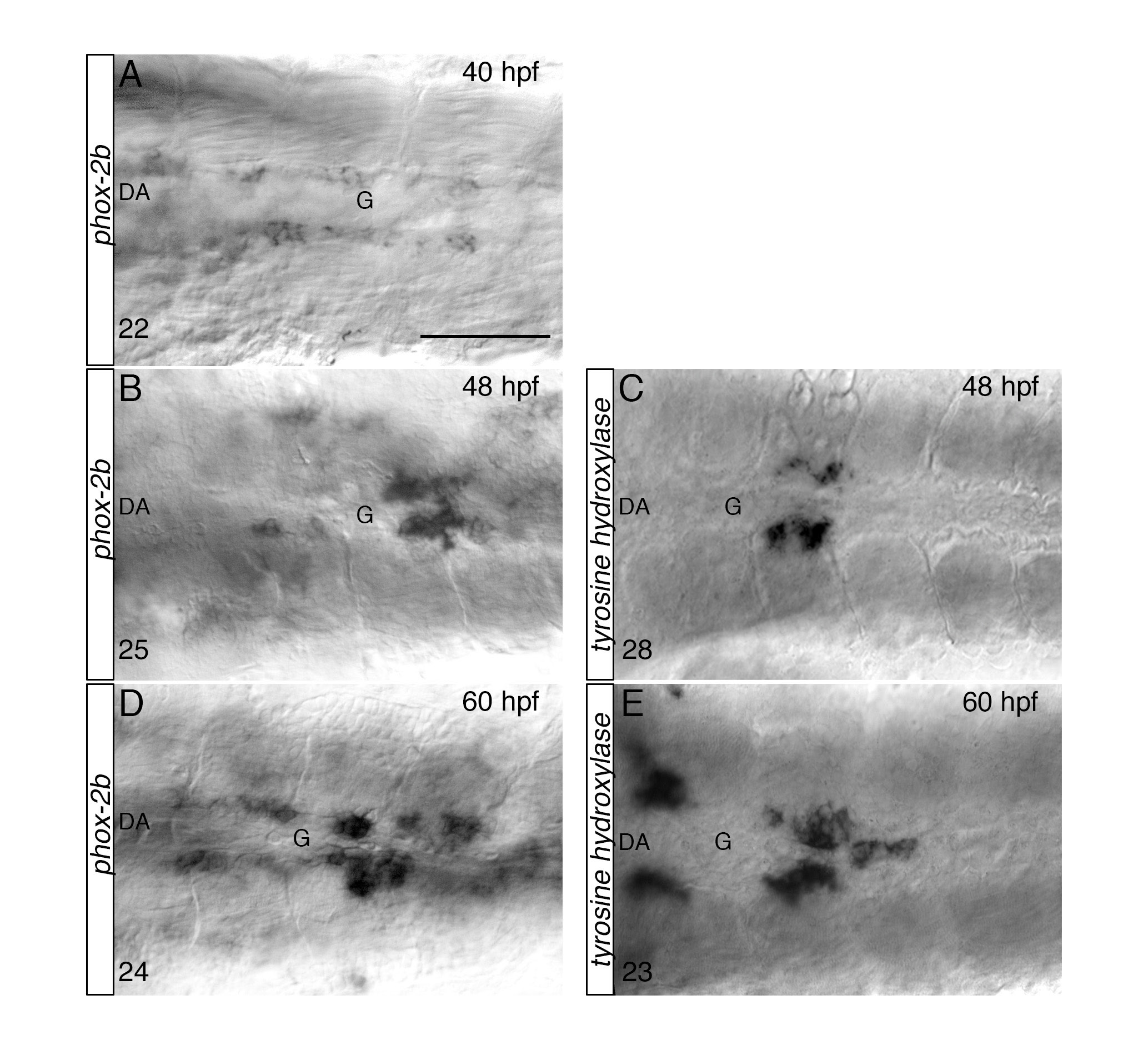
Fig. S2 Stepwise differentiation of SNs next to the DA, related to Figure 2. (AE) Whole-mount in situ hybridizations with antisense riboprobes specific for phox-2b (A, B, D) and tyrosine hydroxylase (th) (C, E) at the indicated time points. Images are ventral views (anterior is to the left) of the region between LDA-DA connection (left) and the glomerular region (G) after dissection of the yolk sac. Phox-2b positive sympathetic precursors are present around the DA at 40 hpf (A), before th+ RNA expressing cells appear at 48 hpf (C). Phox-2b and th expressions patterns expand to more anterior and posterior regions at 60 hpf (D, E). The number of embryos observed is indicated at the bottom left. Scale bar: 75 µm in A-E.
Image
Figure Caption
Acknowledgments
This image is the copyrighted work of the attributed author or publisher, and
ZFIN has permission only to display this image to its users.
Additional permissions should be obtained from the applicable author or publisher of the image.
Full text @ Cell Rep.

