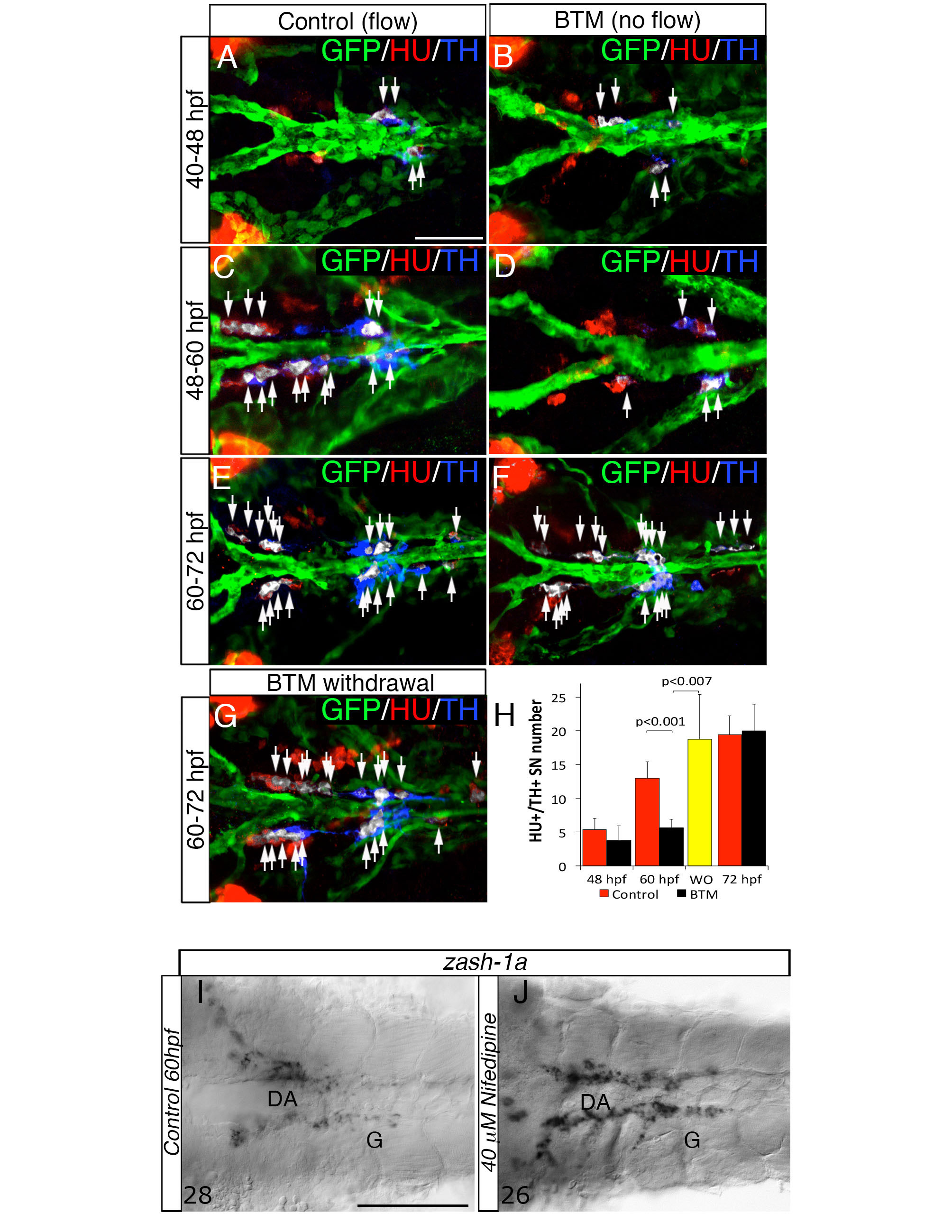
Fig. S3 Blood flow is required at specific stages to induce NA differentiation of SNs, related to Figure 4. (A-F) Dorsal views (anterior to the left) of control- (A, C, E) or 2,3 Butanedione monoxime (BTM)-treated (B, D, F) Tg(kdrl:EGFP)la116 zebrafish embryos. HU+/TH+ SNs are pseudocolored in white and indicated (arrows). BTM was used to stop the blood flow between the stages indicated on the left. Embryos exposed to 20 mM BTM from 48 to 60 hpf (C-D) show severe interruption of the NA differentiation of SNs. BTM treatment from 40 - 48 hpf or from 60 - 72 hpf does not affect the NA differentiation of SNs (A-B, E-F). (G) After BTM exposure between 48 – 60 hpf, the drug was withdrawn and embryos were maintained under control conditions for an additional 12h (60-72 hpf). NA differentiation recovered following BTM withdrawal. (H) Quantitative analysis of HU+/ TH+ SNs in control- (red bars), BTM -treated (black bars) and BTM withdrawal (WO) (yellow bar) embryos. Data were calculated from three independent experiments. N.S., not significant; error bars indicate SD. (I, J) Whole-mount in situ hybridizations with antisense riboprobe specific for zash-1a RNA in control (I) and embryos treated with Nifedipine between 48 and 60 hpf (J). Images are ventral views (anterior is to the left) of the region between LDA-DA connection (left) and the glomerular region (G) after dissection of the yolk sac. Nifedipine did not affect Zash-1a positive sympathetic precursors. Numbers of analyzed embryos are indicated. Scale bars: 50 µm in A-G, 75 µm in I,J.
Image
Figure Caption
Acknowledgments
This image is the copyrighted work of the attributed author or publisher, and
ZFIN has permission only to display this image to its users.
Additional permissions should be obtained from the applicable author or publisher of the image.
Full text @ Cell Rep.

