Fig. 1
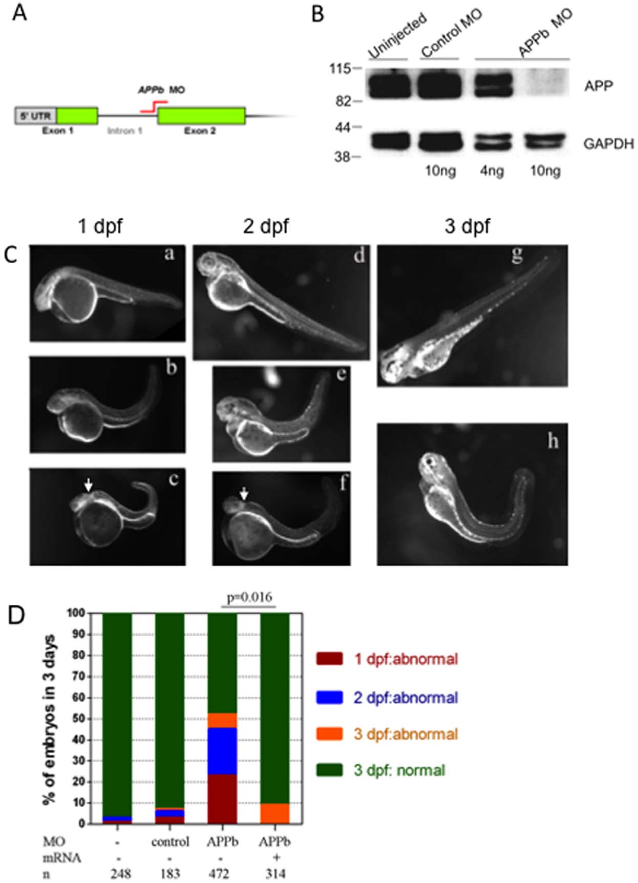
APPb is required for normal zebrafish embryonic development.
(A) Schematic representation of APPb-MO blocking the mRNA splicing site between intron 1 and exon 2 (indicted in red) as used in this study. (B) Western blot analysis of APPb protein in zebrafish embryos at 2 dpf. The lower panel was probed with anti-GAPDH antibody. At 2 dpf, APPb protein migrated as a doublet at 98 KDa with stronger expressions in the un-injected group and control MO group. With the injection of 10 ng of APPb MO per embryo, APPb protein levels were not detected at 2 dpf. With the injection of 4 ng of APPb MO per embryo, weak expression of APPb protein migrating at 98 KDa was observed. (C) Morphological features of control embryos (a, d, g) and APPb morphant embryos (b, c, e, f, h). Lateral views (anterior to the left and dorsal at the top) of zebrafish embryos. The gross anatomical phenotype included a deformed body and a shortened and curved tail. In addition, defects in midbrain patterning were observed (arrows). 1 dpf: a, b, c; 2 dpf: d, e, f; 3 dpf: g, h. (D) APPb mRNA rescues the defective phenotype. There is little effect on normal embryonic development caused by the injection of control morpholino (APPb mis-match MO). Zebrafish embryos injected with 10 ng of APPb-MO expressed abnormal phenotypes at the 1 dpf (red), 2 dpf (blue) and 3 dpf (yellow) developmental stages. Embryos co-injected with 10 ng of APPb-MO and 350 pg APPb mRNA expressed normal phenotypes during embryogenesis. Statistical significance was measured comparing APPb-MO embryos and the co-injected embryos (p = 0.016, p<0.05, in 2-tailed paired t tests).

