Image
Figure Caption
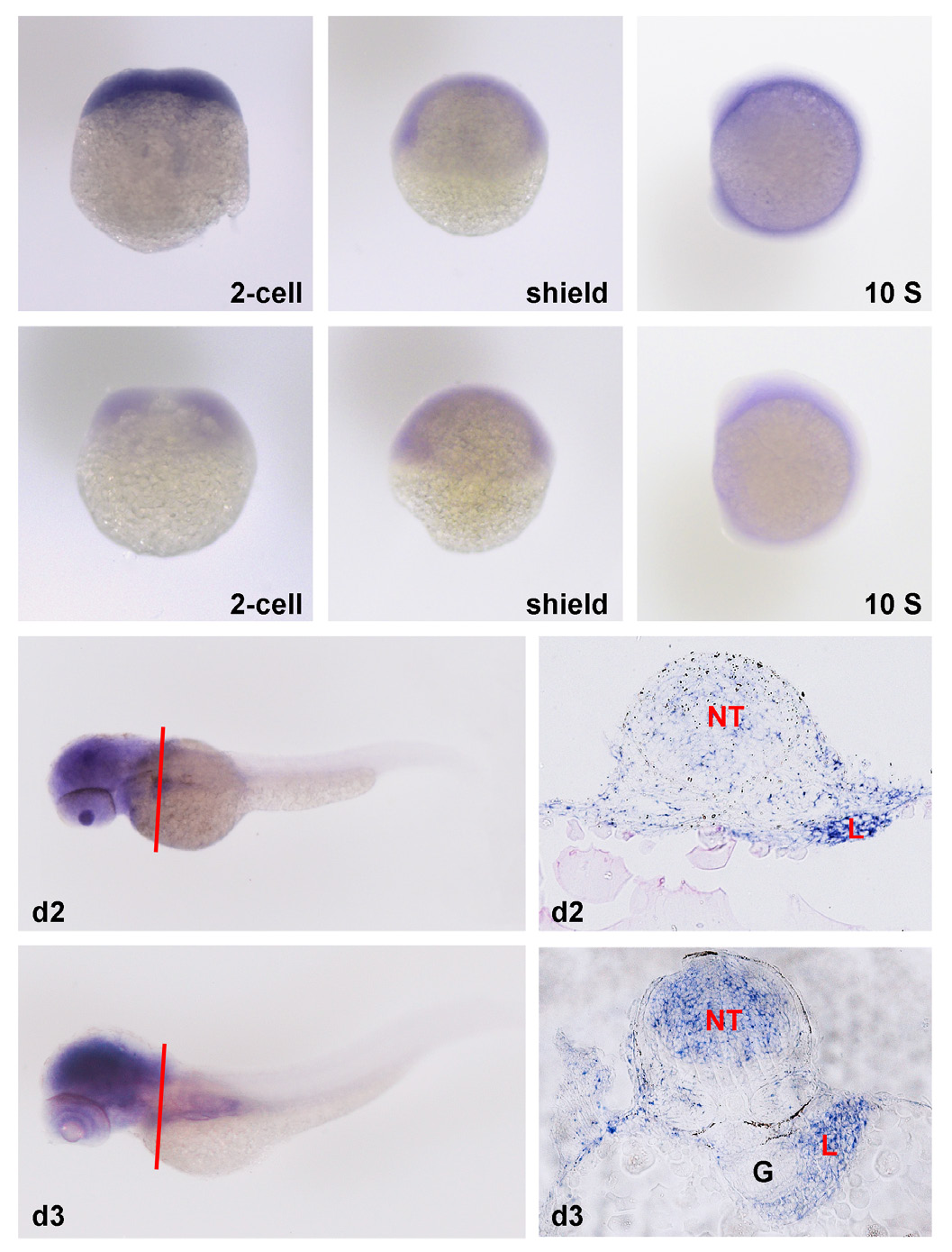
Fig. 1 The embryonic expression pattern of zebrafish zfyve9a. Embryos were hybridized to an antisense probe to zfyve9a (A-C, G-J) or a sense control probe (D-F). (A) Maternal zfyve9a mRNA was detected at 2-cell stage. (B-C) Low level expression of zfyve9a at the shield and 10-somite stages. (G-I) zfyve9a was enriched in the liver at day 2 and 3. It was also highly expressed in the brain and neural tube at day 3. (H) is the cross-section of (G) at the position indicated by the red line. (J) is the cross-section of (I) at the indicated position. NT, neural tube; L, liver; G, gut.
Figure Data
Acknowledgments
This image is the copyrighted work of the attributed author or publisher, and
ZFIN has permission only to display this image to its users.
Additional permissions should be obtained from the applicable author or publisher of the image.
Full text @ Int. J. Dev. Biol.

