
Image
|
Figure Caption
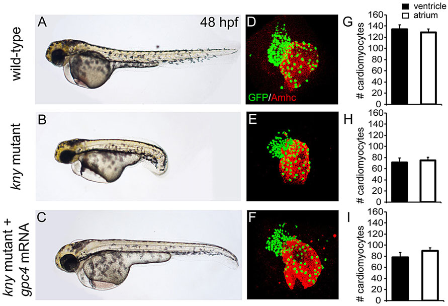
Fig. 1
Reduced cardiomyocyte numbers in kny/gpc4 mutants. (A-C) Lateral view of wild type (WT) (A), kny/gpc4 mutant (B) and kny/gpc4 mutant injected with kny/gpc4 mRNA (C) at 48hpf. (D-F) Confocal images of hearts from Tg(myl7:galFF/UAS:h2a-gfp, kny+/) embryos stained for GFP and Amhc derived from WT (D), kny/gpc4 mutant (E) and kny/gpc4 mutant injected with kny/gpc4 mRNA (F). (G-I) Quantification of number of cardiomyocytes located in either the ventricle or atrium of WT (G), kny/gpc4 mutant (H) and kny/gpc4 mutant injected with kny/gpc4 mRNA (I) at 48hpf (n=3). Results are represented as mean�s.e.m.
Acknowledgments
This image is the copyrighted work of the attributed author or publisher, and
ZFIN has permission only to display this image to its users.
Additional permissions should be obtained from the applicable author or publisher of the image.
Full text @ Development