
Image
|
Figure Caption
Fig. 3
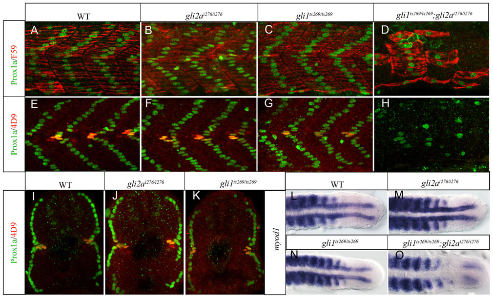
Gli1 and Gli2a act redundantly to pattern the zebrafish myotome.
(A,E) Wild-type (WT); (B,F) gli2ai276; (C,G) gli1ts269 and (D,H) gli1ts269; gli2ai276 24hpf embryos stained with anti Prox1a (green) and either mAb F59 (red: panels A?D) or 4D9 (red: panels E?H) to reveal slow-twitch muscle and MP fibres. (I?K) Transverse sections of embryos shown in panels E?G. (L?O) Dorsal views of the caudal regions of 18ss embryos hybridized with a probe for myod1 transcript. (L) WT; (M) gli2ai276; (N) gli1ts269 (O) gli1ts269;gli2ai276.
Figure Data
Acknowledgments
This image is the copyrighted work of the attributed author or publisher, and
ZFIN has permission only to display this image to its users.
Additional permissions should be obtained from the applicable author or publisher of the image.
Full text @ Biol. Open