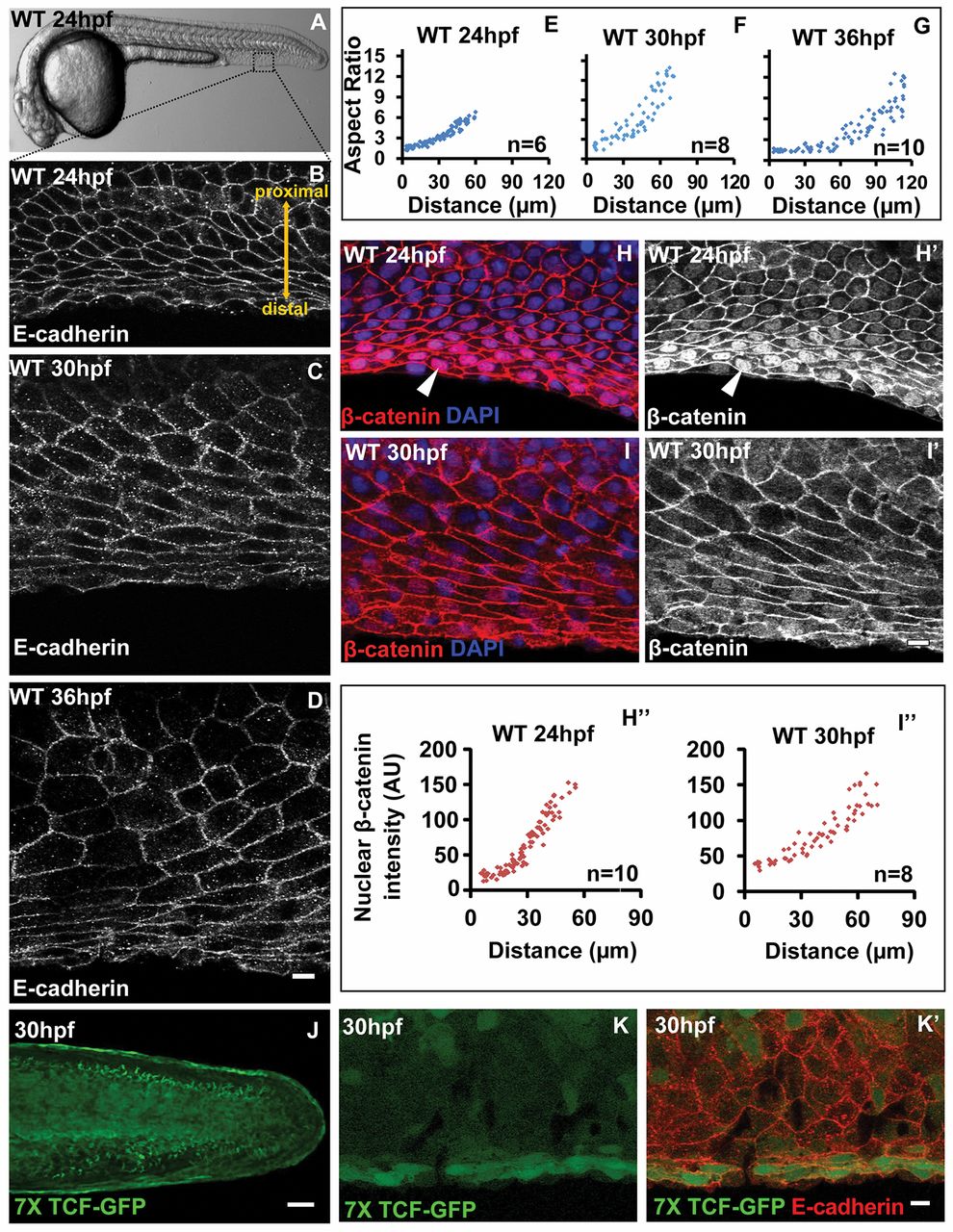
Fig. 1 Correlation between cellular pattern and Wnt signalling gradient in the median fin fold epithelium. (A) Bright-field image of 24 hpf wild-type zebrafish embryo. The dotted box represents the region of the fin epithelium imaged by confocal microscopy. (B-G) E-cadherin staining and aspect ratio plots of median fin epithelial cells at 24 (B,E), 30 (C,F) and 36 hpf (D,G) in wild-type embryos. Aspect ratios are plotted against the distance of the cells from the base of the fin fold along PD axis. (H-I′) β-Catenin-DAPI overlays (H,I) and β-catenin staining (H′,I′) and its quantification (H",I") in median fin epithelium at 24 hpf (H-H′) and 30 hpf (I-I′). (J-K′) Tg(7XTCF-Xia.Siam:GFP)ia4 line showing GFP expression in the distal cells of the median fin at lower magnification (J) and at higher magnification (K,K′) along with E-cadherin staining. Arrowheads in H,H′ indicate nuclear β-catenin. Scale bars: 10 μm B-D,H-I′,K,K′ ; 50 μm in J. AU, arbitrary units.
Image
Figure Caption
Figure Data
Acknowledgments
This image is the copyrighted work of the attributed author or publisher, and
ZFIN has permission only to display this image to its users.
Additional permissions should be obtained from the applicable author or publisher of the image.
Full text @ Development

