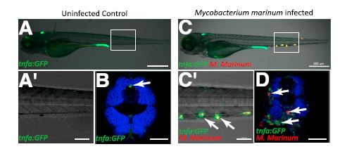
Fig. S3 M. marinum infection induces expression of TgBAC(tnfa:GFP) in WT larvae. (A and A′) Uninfected control TgBAC(tnfa:GFP) 120 hpf larvae at low magnification (A) and higher magnification of boxed area (A′). (B) Confocal image of a cross-section of an uninfected 120 hpf WT larva. Phalloidin, blue. Arrow points to basal TgBAC(tnfa:GFP) expression. (C and C′) M. marinum-infected (red) 120 hpf TgBAC(tnfa:GFP) larva at low magnification (C) and higher magnification of boxed area (C′). Arrows point to colocalization of TgBAC(tnfa:GFP) positive cells with M. marinum. (D) Cross-section through M. marinum-infected (red) 120 hpf TgBAC(tnfa:GFP) larva. Phalloidin, blue. Arrows point to TgBAC(tnfa:GFP) positive granulomas surrounding M. marinum bacteria. (Scale bars: A and C, 500 Ám; A′ and C′, 100 Ám; B and D, 50 Ám.)
Image
Figure Caption
Acknowledgments
This image is the copyrighted work of the attributed author or publisher, and
ZFIN has permission only to display this image to its users.
Additional permissions should be obtained from the applicable author or publisher of the image.
Full text @ Proc. Natl. Acad. Sci. USA

