
|
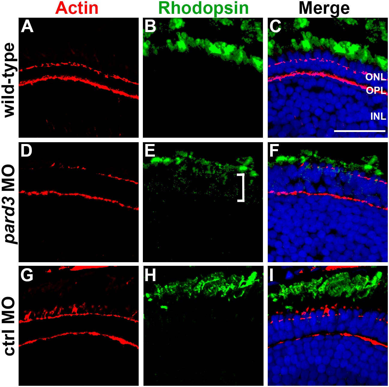
Fig. 7 Rhodopsin is mislocalized in pard3 morphants. (A) Immunohistochemistry was performed on transverse cryosections through the retina of 5 dpf wild-type larvae. Left most panels show phalloidin staining to label actin (red). Rhodopsin immunoreactivity (green) is shown in middle panels. (D–F) In pard3 morphants, outer segments appear slightly shorter and opsin staining within the inner segment is observed (bracket). (G–I) Rhodopsin localizes normally in control morphants. Sections were also counterstained with DAPI (blue). ONL = outer nuclear layer; OPL = outer plexiform layer; INL = inner nuclear layer. Scale bar = 20 μm.