
|
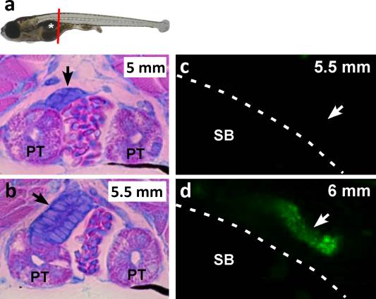
Fig. 1
Formation of the first mesonephric nephron. A: A cross section of a 5 mm larva stained with methylene blue and basic fuchsin shows a basophilic cluster sitting on top of the PT (arrow) at the caudal end of the swim bladder (white asterisk). The red line indicates the region of the cross sections for A-B. B: A cross section of a 5.5 mm larva stained similarly to A shows a nascent basophilic tubule making contact with the PT (arrow), but its lumen has not yet fused with the PT lumen. C: Injection of 40 kDa dextran-FITC into a 5.5 mm larva indicates that the nascent tubule is not yet functional and did not accumulate the fluorescent tracer (arrow). D: The tubule of a 6 mm larva did accumulate the fluorescent tracer (arrow), indicating that the first mesonephric nephron is functional at the 6 mm stage. PT: pronephric tubule; SB: swim bladder; dashed lines demarcate the swim bladder from the pronephric tubule.