
|
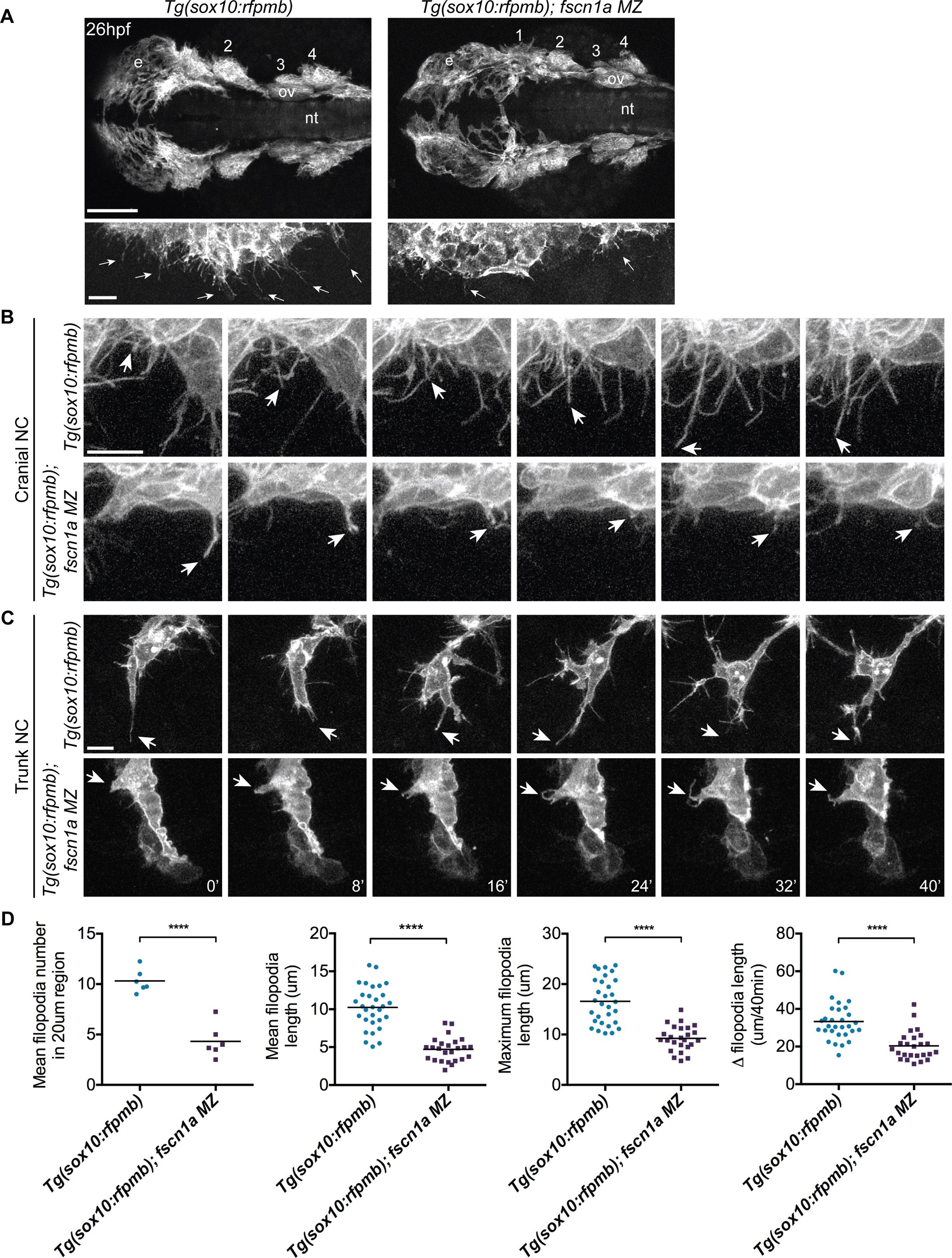
Fig. 3 fscn1a is required for filopodia formation in NC cells.
(A) Dorsal cranial views of cranial NC streams in 26 hpf Tg(sox10:rfpmb) and Tg(sox10:rfpmb); fscn1a MZ embryos, anterior is to the left. Numbers correspond to pharyngeal arches, e = eye, ov = otic vesicle, nt = neural tube, scale bar = 100µm. Leading edge of NC stream 1 is enlarged in right panel, scale bar = 10 µm. Arrows highlight filopodia at leading edge of NC stream 1. (B-C) Time lapse confocal images of filopodia at the leading edge of NC stream 2 (B) or of leading cells in trunk NC streams (C) in live Tg(sox10:rfpmb) and Tg(sox10:rfpmb); fscn1a MZ embryos at 26 hpf. Arrows highlight tips of single filopodia throughout the time lapses. Scale bar = 10 µm. (D) Quantitation of filopodia number, mean and maximum filopodia length, and change in filopodia length in Tg(sox10:rfpmb) and Tg(sox10:rfpmb); fscn1a MZ cranial NC (n = 6 embryos and 30 filopodia each for Tg(sox10:rfpmb) and Tg(sox10:rfpmb); fscn1a MZ, ****p<0.0001).