Fig. 4
Laminin-211 Concentration Modulates In Vitro Myelination
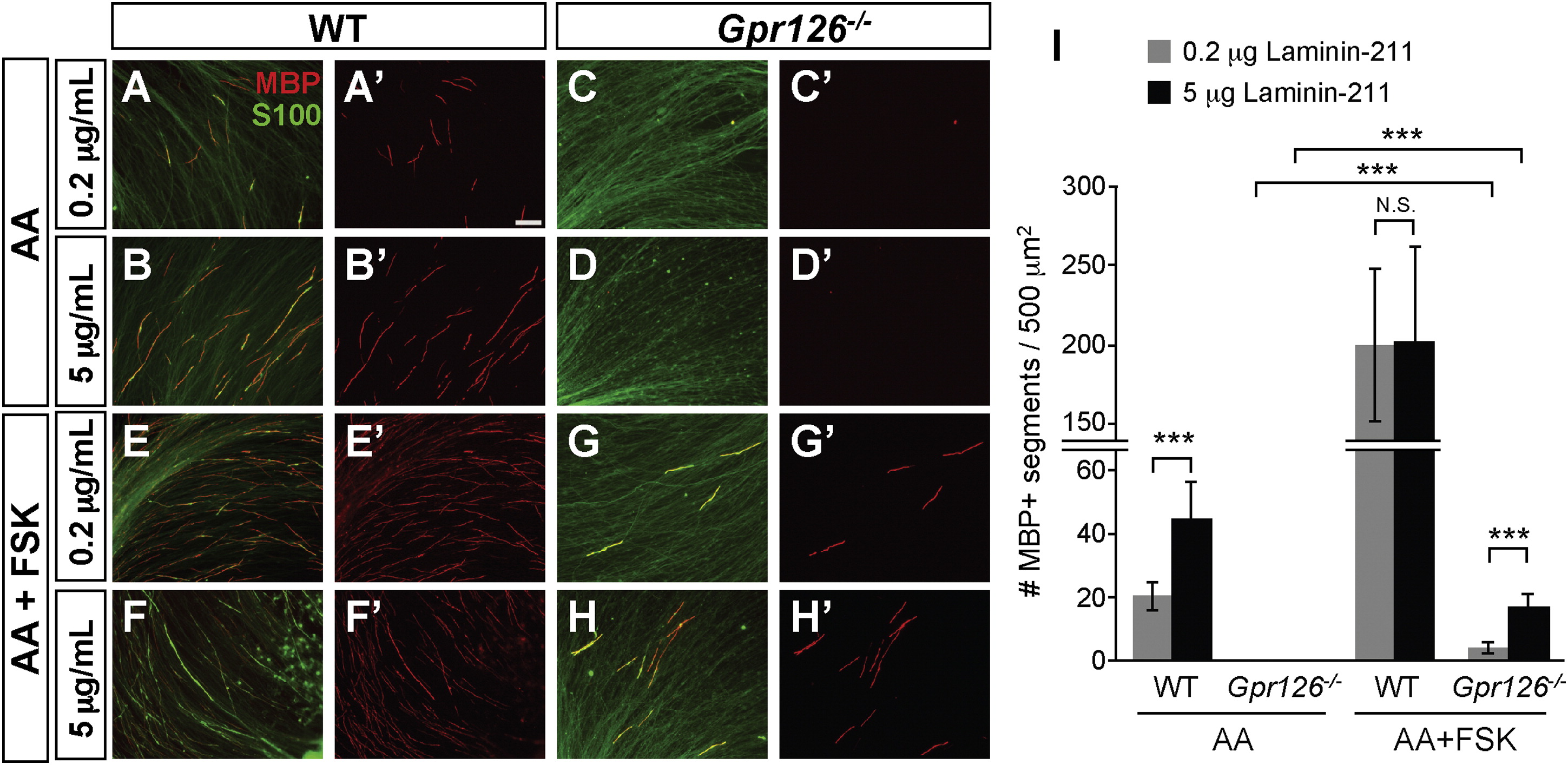
(A?H) In vitro myelination cultures from WT or Gpr126/ DRGs on plates coated with poly-D-lysine + 0.2 or 5 µg/ml Laminin-211. Cultures were immunostained with MBP (red) and S100 (green). The scale bar represents 100 µm.
(A and B) Three weeks after ascorbic acid (AA) addition to WT, more MBP+ immunostaining is observed on 5 µg/ml Laminin-211 than 0.2 µg/ml 3 weeks after AA addition.
(C and D) MBP+ immunostaining is not observed in Gpr126/ SCs at any Laminin-211 concentration 3 weeks after AA addition.
(E and F) Addition of FSK leads to MBP potentiation in WT SCs on both 0.2 and 5 µg/ml Laminin-211 relative to AA alone.
(G and H) Gpr126/ SCs have low levels of MBP in the presence of FSK and show concentration dependence for Laminin-211.
(I) Quantification of myelination cultures. Bars represent the average number of MBP-positive segments per 500 µm2 ± SD. p < 0.001, Student?s t test; N.S., no significant difference.

