
|
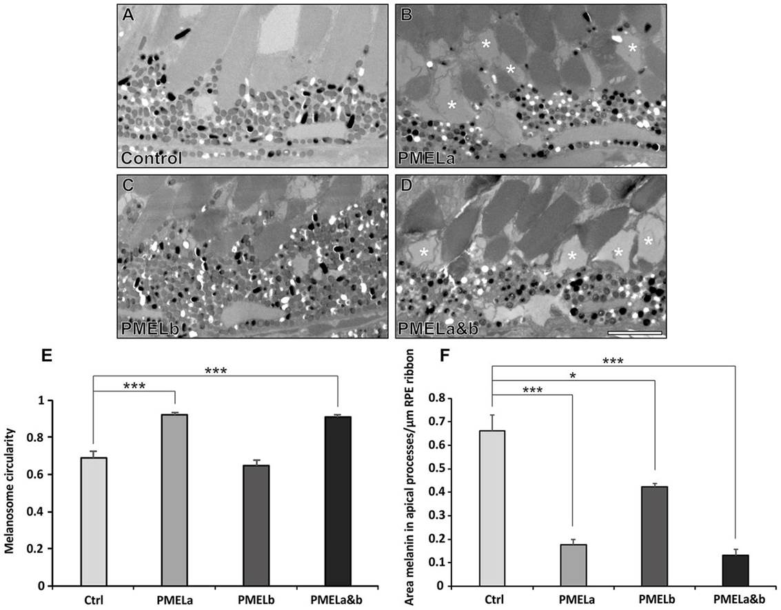
Fig. 6
PMELa MO prevents melanosome movement into the apical processes and has a profound effect on photoreceptor morphology. (A?D) Electron micrographs showing the effect on melanosome positioning in the apical processes at 5dpf for zebrafish injected with control (A), PMELa (B), PMELb (C) and PMELa&b MOs (D). (B,D) PMELa and PMELa&b MOs resulted in missing or disrupted outer segments as shown by the asterisks. Scale bars: 4�m. (E) Photoreceptors in fish injected with PMELa and PMELa&b MOs had significantly fewer cylindrical melanosomes at 5dpf. Ctrl, control. (F) PMELa and PMELa&b MOs resulted in a reduced area of melanin (thus fewer melanosomes) in the apical processes. Results show the mean�s.e.m.; *P<0.05, ***P<0.001 (Student′s t-test).