
Image
|
Figure Caption
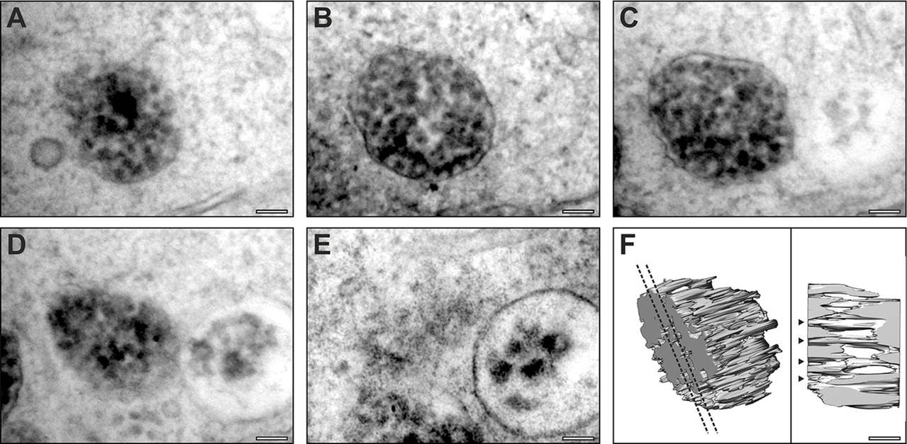
Fig. 3
Serial section electron microscopy analysis of tyrosinase MO-treated RPE reveals immature melanosomes that contain fibrils. (A–E) Serial section micrographs of the same immature melanosome in an RPE cell from a tyrosinase MO-treated zebrafish at 5dpf. (F) 3D rendering of the micrograph data reveals fibrils running through the melanosome. Dotted lines indicate the position of the slice shown in the right-hand panel; arrowheads indicate fibrils running through the entire thickness of the melanosome. Scale bars: 100 nm.
Figure Data
Acknowledgments
This image is the copyrighted work of the attributed author or publisher, and
ZFIN has permission only to display this image to its users.
Additional permissions should be obtained from the applicable author or publisher of the image.
Full text @ J. Cell Sci.