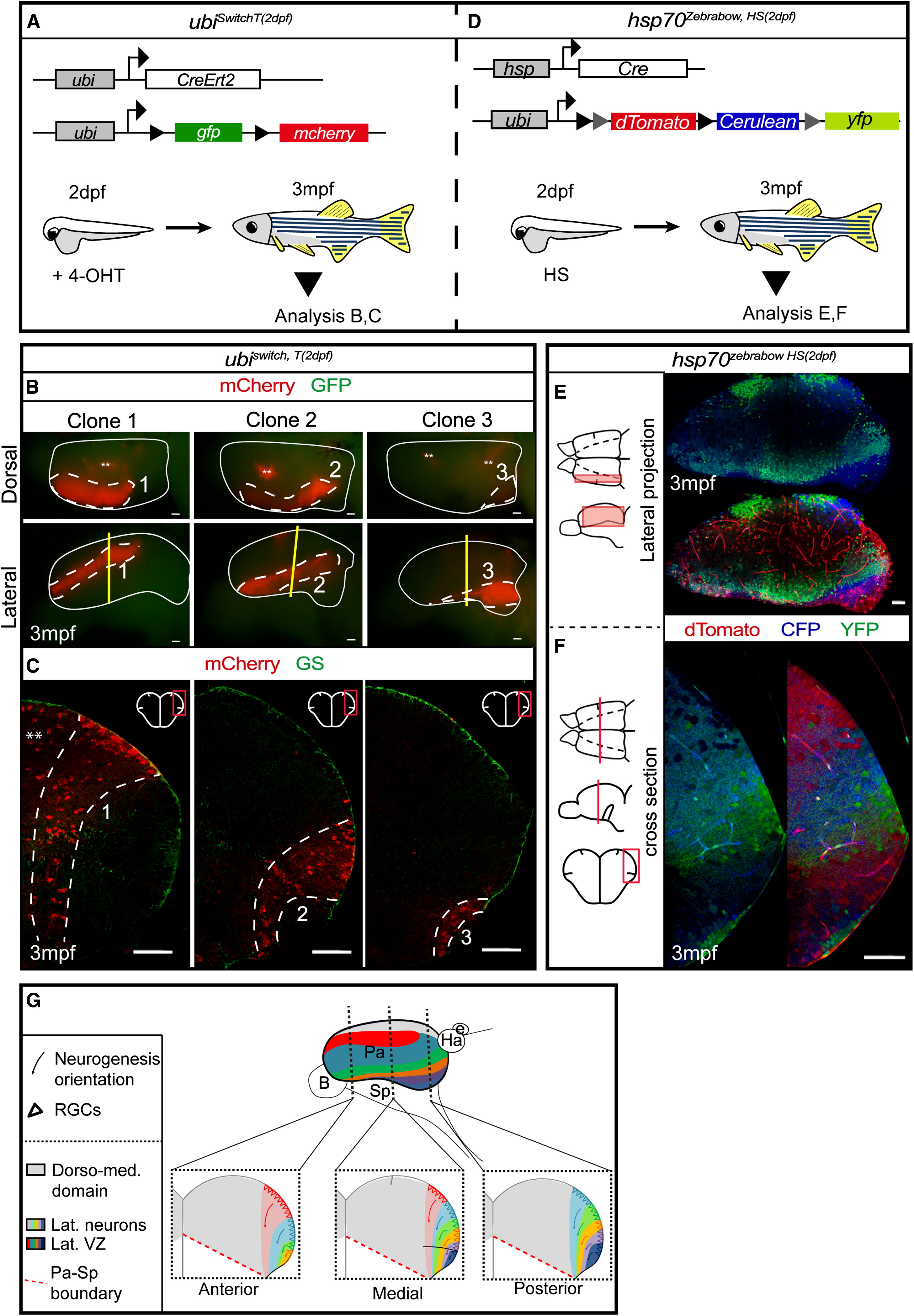
Fig. 2
A Restricted Number of Progenitors at 2 dpf Generate the Lateral aNSCs Following Massive Postembryonic Amplification
(A) Experimental design to analyze the morphology of aNSCs polyclones generated from embryonic progenitors in the lateral pallium.
(B) Dorsal (top) and lateral views (bottom) of ubiswitch,T(2 dpf) telencephali (whole mount of one hemisphere, anterior left). Representative lateral clone types 1, 2, and 3 are shown (dotted lines). Asterisks highlight medial pallial clones.
(C) Cross-sections of the telencephali shown in (B; section plane: yellow) and stained as indicated.
(D) Experimental design to map the adult fate of individual early pallial progenitors in single brains.
(E) Lateral projections of the adult pallium (whole-mount) in hsp70zebrabow,HS(2 dpf) fish showing expression of CFP/YFP (top) or CFP/YFP/dTomato (bottom).
(F) Cross-section of the telencephalon of hsp70zebrabow,HS(2 dpf) adults showing expression of CFP/YFP (left) or CFP/YFP/dTomato (right).
(G) Scheme depicting the typical morphology of lateral pallial clones after a recombination as in (A) and (D), and cross-sections at anterior, medial, and posterior levels. Triangles represent the RGCs, colored domains represent the neurons generated from these progenitors, and arrows represent the progression of neurogenesis.
See also Figure S2.
Reprinted from Developmental Cell, 30(2), Dirian, L., Galant, S., Coolen, M., Chen, W., Bedu, S., Houart, C., Bally-Cuif, L., Foucher, I., Spatial Regionalization and Heterochrony in the Formation of Adult Pallial Neural Stem Cells, 123-36, Copyright (2014) with permission from Elsevier. Full text @ Dev. Cell

