
|
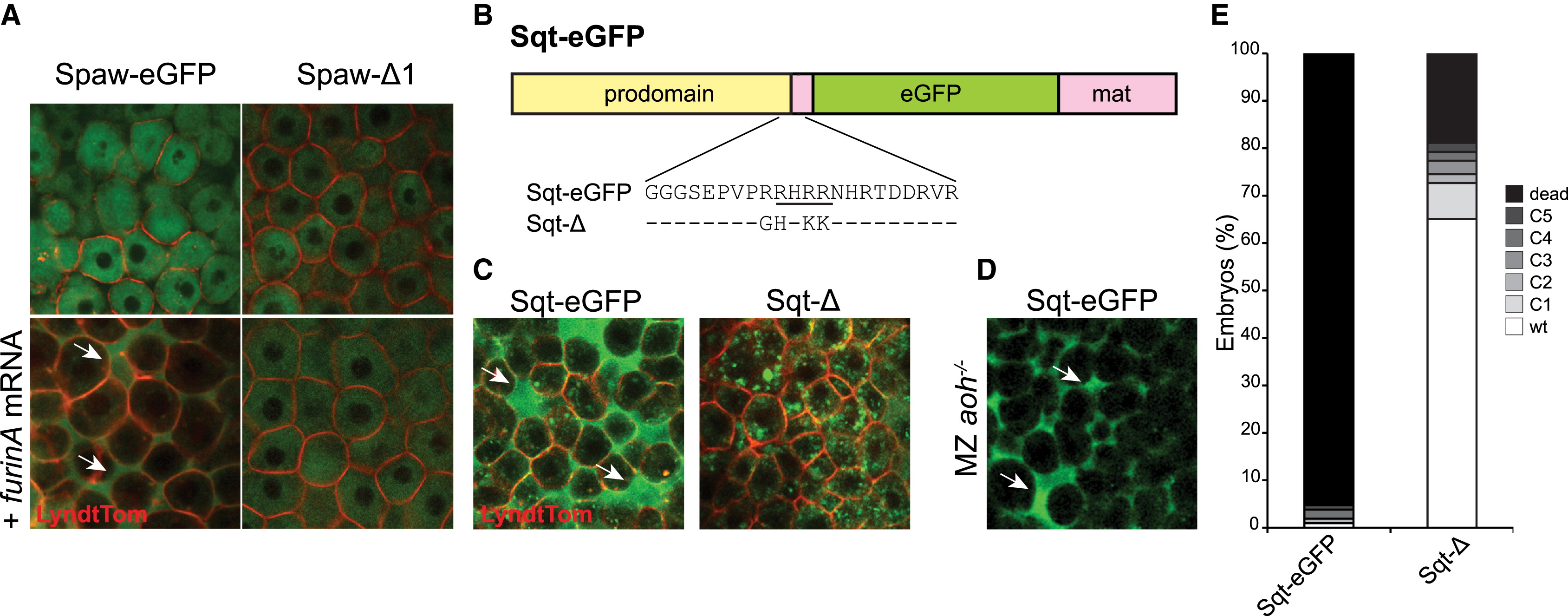
Fig. 3
FurinA-Mediated Processing of Spaw Specifically Correlates with Extracellular Localization of Spaw
(A) Embryos were injected at the one-cell stage with 50 pg of Spaw-EGFP mRNA or Spaw-Δ1 mRNA together with a membrane-bound tdTomato fusion protein and imaged at sphere stage (4 hpf). Addition of FurinA mRNA (30 pg) to the injection mix resulted in an increased extracellular localization of Spaw-EGFP, but not Spaw-?1.
(B) Schematic representation of the Sqt-EGFP fusion protein (M�ller et al., 2012). The Furin recognition site is underlined in the detail of the sequence. The changes in amino acids in the noncleavable form Sqt-? are detailed.
(C) Removal of the Furin cleavage site (Sqt-?) resulted in reduced extracellular localization of Sqt-EGFP. mRNA injections and imaging were carried out identically to (A).
(D) MZaoh mutant embryos were injected at the one-cell stage with 50 pg Sqt-EGFP mRNA and imaged at sphere stage (4 hpf). Arrows indicate extracellular localization of the Spaw-EGFP and Sqt-EGFP fusion proteins respectively in (A), (C), and (D).
(E) Quantification of the dorsalization phenotypes induced by injection of 1 pg Sqt-EGFP and Sqt-?. The embryos were scored as described in Figure 1I.
Reprinted from Developmental Cell, 32(5), Tessadori, F., No�l, E.S., Rens, E.G., Magliozzi, R., Evers-van Gogh, I.J., Guardavaccaro, D., Merks, R.M., Bakkers, J., Nodal Signaling Range Is Regulated by Proprotein Convertase-Mediated Maturation, 631-9, Copyright (2015) with permission from Elsevier. Full text @ Dev. Cell