
|
Fig. 6
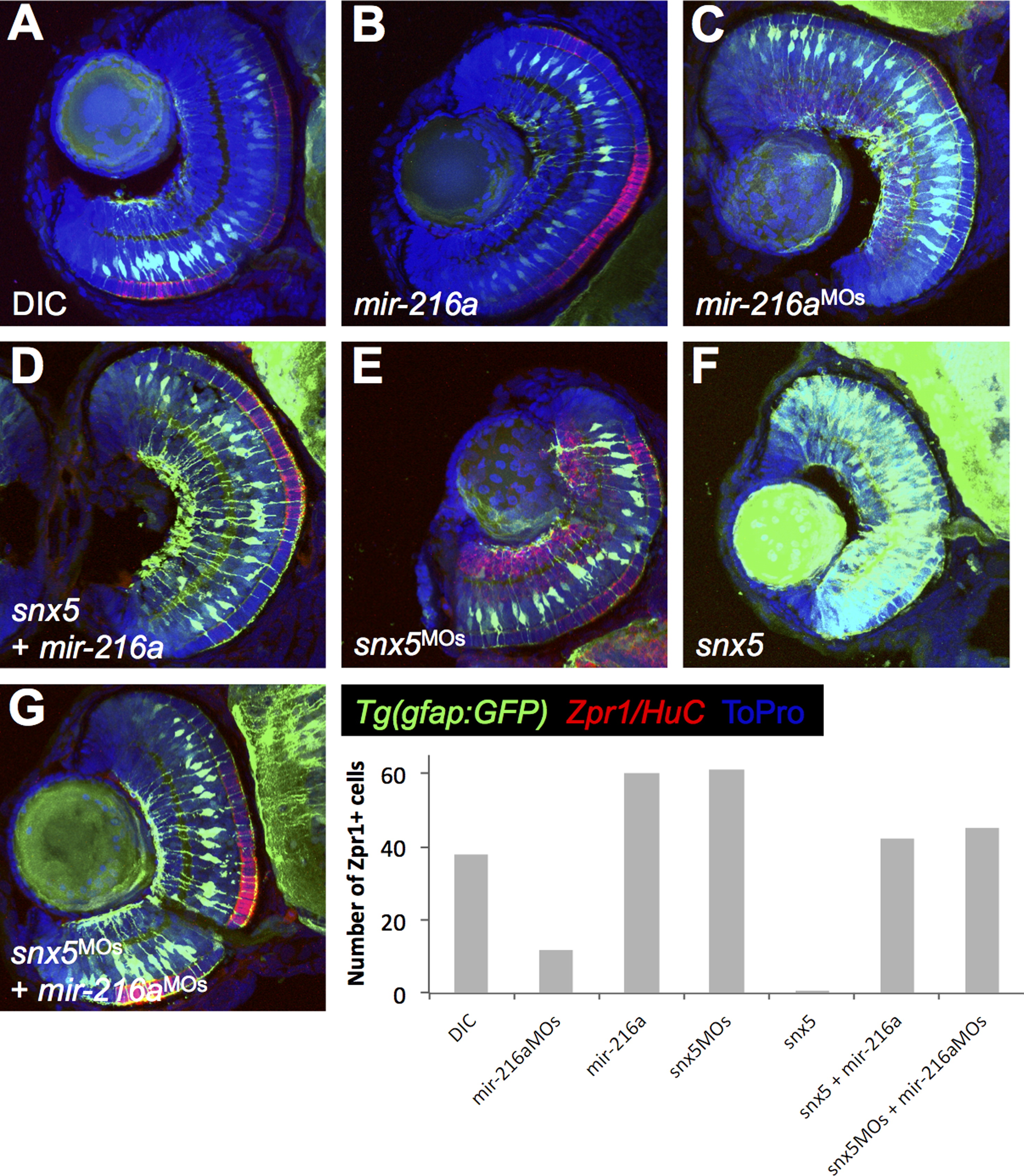
Inverse correlation between MG numbers and cone photoreceptor staining. Tg(gfap:gfp) embryos were injected with dye control (DIC; A), miR-216a (B), miR-216aMOs (C), snx5MOs (E), or snx5 mRNA (F) at the 1-cell stage, fixed at 65 hpf, and transverse sections of developing retinas were obtained. Immunohistochemistry was performed using antibodies to identify cone photoreceptors in the outer nuclear layer (Zpr-1) or amacrine/ganglion cells in the inner nuclear layer and the ganglion cell layer (HuC). Changes in M�ller glia cell numbers led to consistent changes in cone photoreceptor numbers. Zpr-1 staining increased in embryos injected with mir-216a or snx5MOs and decreased in embryos injected with mir-216aMOs or snx5 compared to embryos injected with dye. Partial rescue of Zpr-1 levels is shown in (D) and (G) where embryos were co-injected with combinations of either snx5 and miR-216a (D) or snx5MOs and miR-216aMOs (G). Amacrine and ganglion cell numbers demonstrated similar, though less striking and less consistent changes compared to cone photoreceptors. Nuclei were marked by staining with To-Pro.
Reprinted from Developmental Biology, 400(1), Olena, A.F., Rao, M.B., Thatcher, E.J., Wu, S.Y., Patton, J.G., miR-216a regulates snx5, a novel notch signaling pathway component, during zebrafish retinal development, 72-81, Copyright (2015) with permission from Elsevier. Full text @ Dev. Biol.