
|
Fig. 3
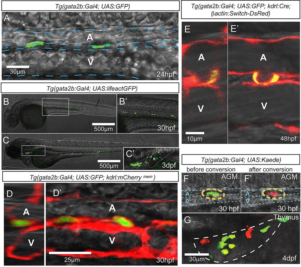
Tg(gata2b:Gal4) marks hemogenic endothelial cells. Imaging is by confocal microscopy. (A) GFP+ endothelial cells in the trunk region of a gata2b:Gal4;UAS:GFP embryo at 24hpf. (B-C′) gata2b:Gal4;UAS:lifeactGFP at 30hpf and 3dpf. (C′) An enlarged view of C, with thymus and kidney highlighted. (D,D′) Transverse (D) and lateral (D′) views of the trunk vasculature of 30hpf gata2b:Gal4;UAS:GFP;kdrl:mCherrymem. (E,E′) Transverse (E) and lateral (E′) views of a 48hpf gata2b:Gal4;UAS:GFP;kdrl:Cre;actB2:LoxP-STOP-LoxP-DsRedEx embryo showing a GFP+ cell undergoing EHT. (F,F′) A representative Kaede+ cell in the trunk of a gata2b:Gal4;UAS:Kaede embryo before (F) and after (F′) photoconversion. Blue dashed lines indicate the approximate location of DA and PCV. (G) Thymus of a 4dpf gata2b:Gal4;UAS:Kaede embryo that experienced photoconversion of a single aortic gata2b+ cell in the trunk at 46hpf. White dashed line highlights the thymus region. T, thymus; K, kidney; A, aorta; V, vein.