Image
Figure Caption
Fig. 6, S1
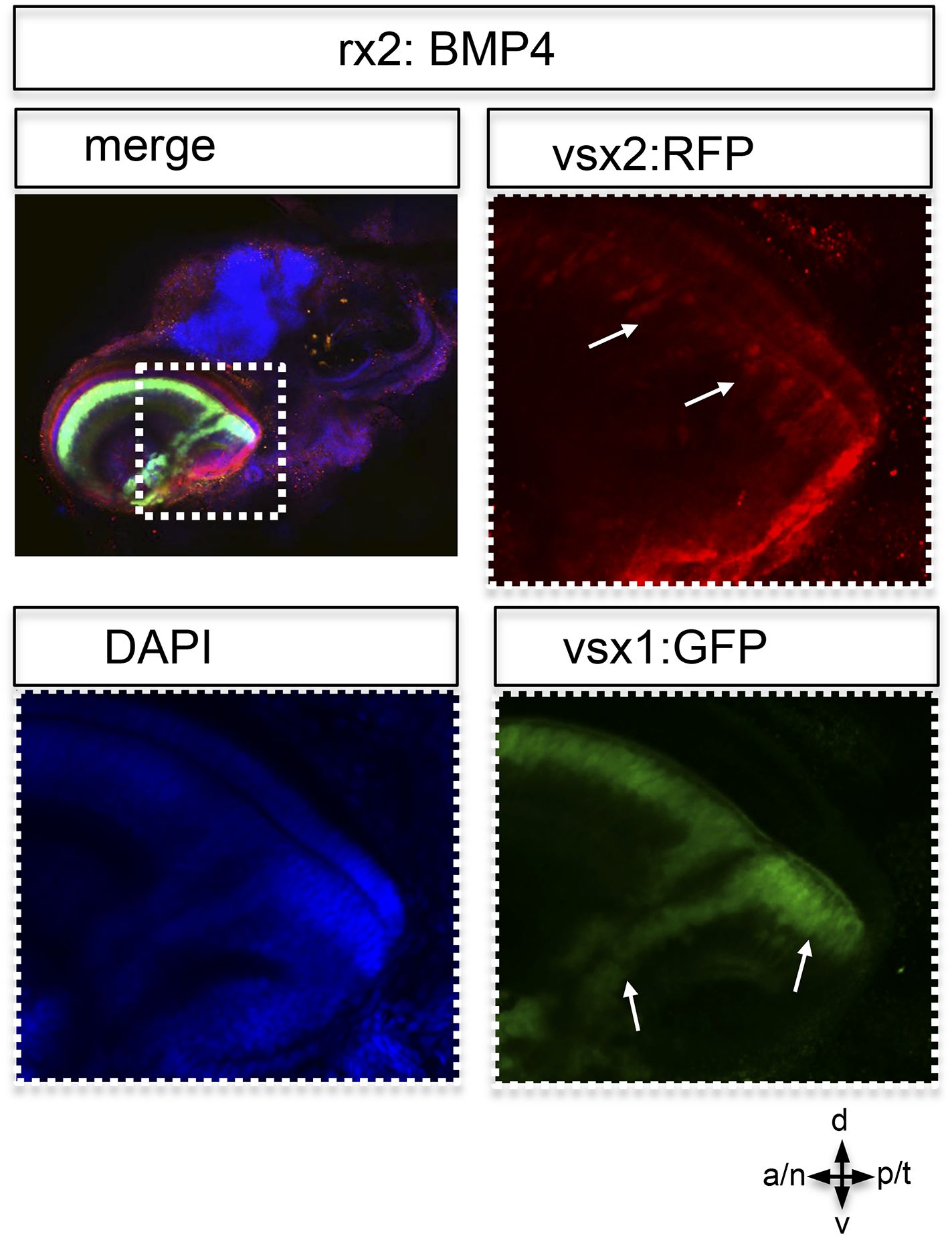
Postembryonic eye development of rx2::BMP4 hatchlings.
Although the temporal optic cup is largely malformed and folded it can be seen clearly, that vsx1 as well as vsx2 transgenes (intensified by wholemount immunohistochemistry) are expressed in the folded epithelium (arrows). This indicates at least a partial correct differentiation into neuroretinal tissue.
Acknowledgments
This image is the copyrighted work of the attributed author or publisher, and
ZFIN has permission only to display this image to its users.
Additional permissions should be obtained from the applicable author or publisher of the image.
Full text @ Elife

