
|
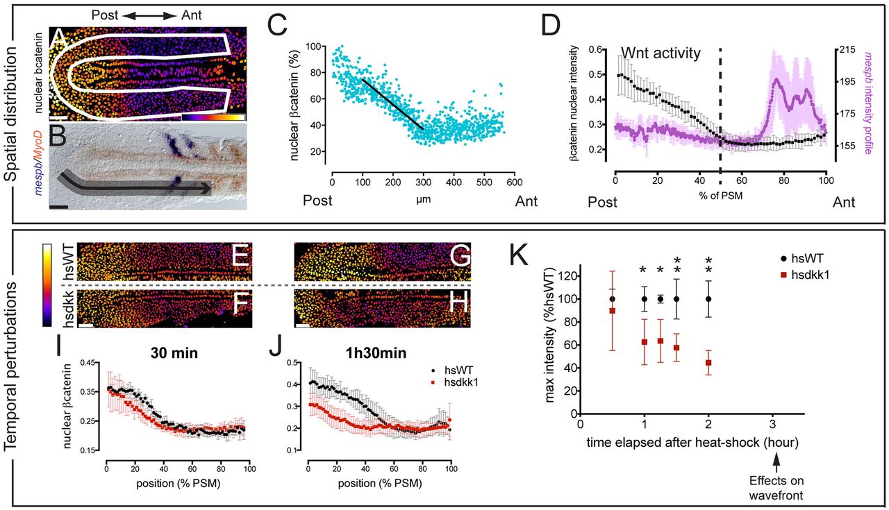
Fig. 4 A posterior gradient of Wnt activity is reduced (A,B,E-H) Dorsal view of flat-mounted non-heat shocked (A,B), hsWT (E,G) or hsdkk1 (F,H) PSM at nine (G,H), ten (A,E,F) or 12 (B) somites. Embryos were fixed 30 minutes (E,F) or 1.5 hours (G,H) phs. (A) Fluorescence intensity of β-catenin staining after applying a nuclear mask, displayed using false color. (B) Whole-mount in situ hybridization with mespb (blue) and myoD (red) probes. (C,D,I,J) Nuclear intensity of β-catenin staining (C, percentage of maximum; D,I,J, grayscale values) along the PSM length for individual cells, for one embryo (C) or averaged within 1.5% bins for several embryos (D,I,J). Error bars reflect variation between embryos. The average mespb intensity profile is in purple in D. (K) Maximum intensity nuclear β-catenin normalized to hsWT dynamic range, with embryos fixed at successive time points. Mean � s.d. *P<0.01, **P<0.001. Scale bars: 50 μm. See also supplementary material Figs S3, S4.