
|
Fig. 6
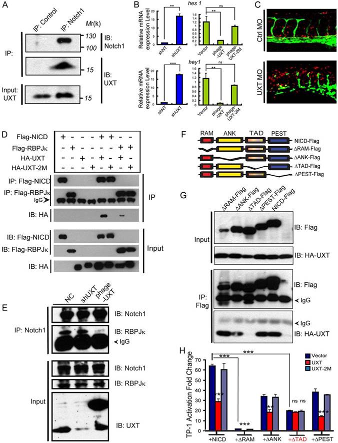
UXT interacts with NICD endogenously through the Notch TAD domain. (A) UXT interacts with NICD endogenously. HUVEC cell lysates were immunoprecipitated (IP) with an anti-Notch1 antibody or control IgG antibody. The immunoprecipitates were immunoblotted (IB) with the indicated antibodies. (B) UXT directly attenuates Notch-targeted genes in endothelial cells. The relative expression level of hes1 and hey1 were measured by RT-PCR. The data were normalized on the basis of the corresponding input control, and are presented as the mean�s.e.m. (at least three independent experiments). (C) UXT attenuates Notch signaling in vivo. Confocal images of Tg(TP1:mCherry; fli1:EGFP)y1 fish injected with 4ng of control MO or 4ng of UXT MO. (D) UXT interacts specifically with NICD. HA-UXT or HA-UXT-2M was co-transfected with Flag-NICD or Flag-RBP-JK. Cell lysates were subjected to an immunoprecipitation assay using the anti-Flag antibody, followed by western blot analysis with anti-HA and anti-Flag antibodies. (E) UXT impairs the interaction between NICD and RBP-JK. HUVECs were treated with shUXT or phage-UXT-Flag. The endogenous NICD was immunoprecipitated with the anti-Notch1 antibody, and the immunoprecipitates were probed with the anti-RBP-JK antibody. (F) Schematic representation of the NICD deletion constructs used in the following experiments. (G) The TAD domain of NICD mediates its interaction with UXT. HA-UXT was co-transfected with ?RAM-Flag, ?ANK-Flag, ?TAD-Flag, ?PEST-Flag or NICD-Flag. Cell lysates were subjected to an immunoprecipitation assay using an anti-Flag antibody. (H) UXT directly impaired Notch signaling. The indicated plasmids were transfected into Cos-7 cells together with TP-1 reporter plasmids, in the presence of NICD or the deletion mutants. Data are presented as the mean�s.e.m. (n=3); **P<0.01; ***P<0.001; ns, non-significant versus the corresponding control or as indicated.