Fig. S4
Mechanical Modeling of Cell Shape Distributions, Related to Figure 4
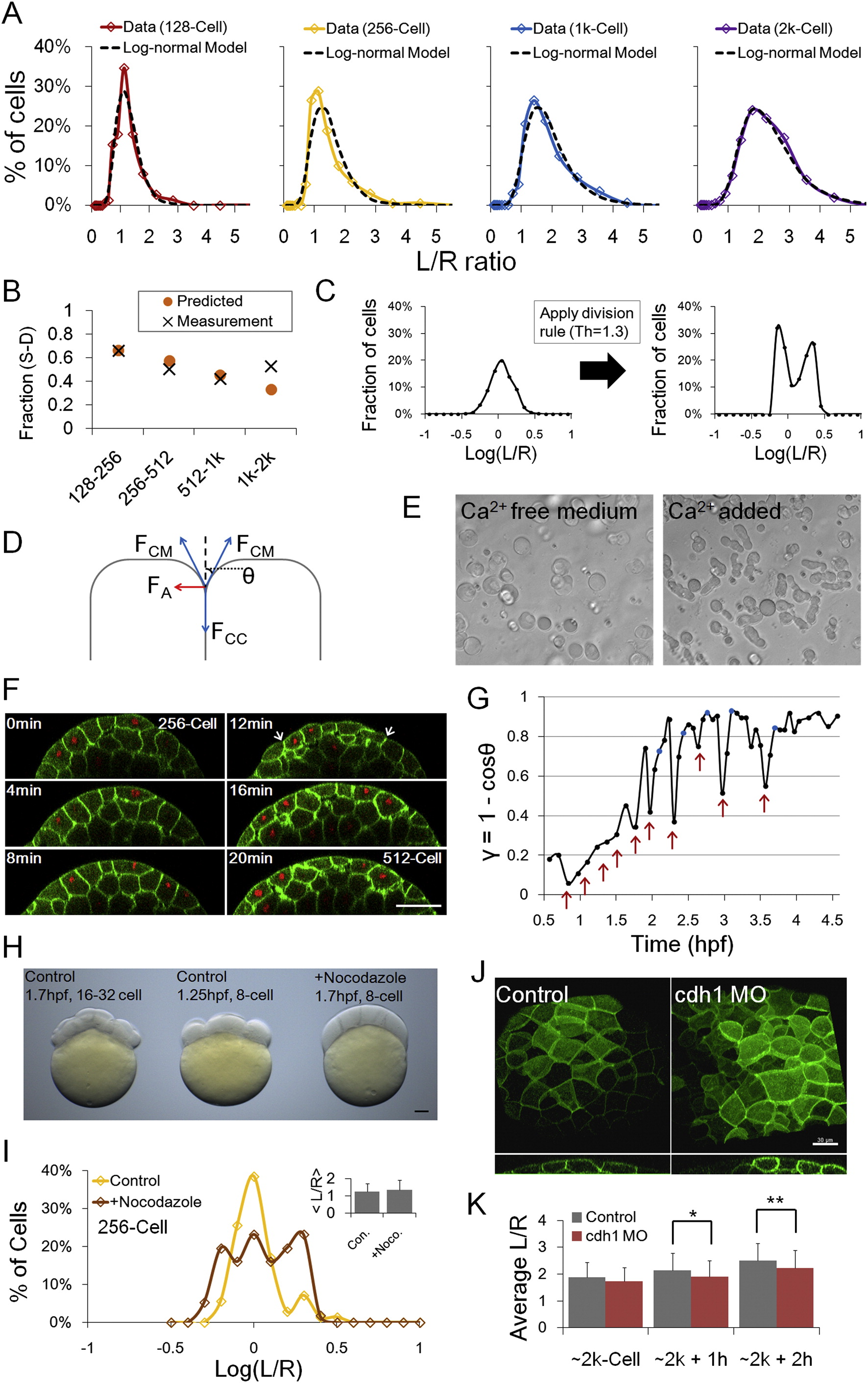
(A) Log-normal fit of data (from Figure 1E) as in Figure 4A. See also Data S1, Text 6.
(B) Predicted S-D division ratio using log-normal fit in (A) and the division rule (Figure 3G).
(C) Predicted L/R distribution using log-normal fit and the division rule. A 2-peak distribution follows when cell shape changes after divisions are not considered.
(D) Local forces that mediate cell shapes. FCM: cell-medium surface tension, FCC: cell-cell surface tension, FA: adhesion force, ?: transition point angle (Maître et al., 2012). See also Data S1, Text 7.
(E) Dissociated cells in Ca2+ free medium round up as spheres under cortical tension; adding Ca2+ allows them to recover adhesion to attach to dish bottom or other cells and deform from sphere.
(F) ? changes during the cell cycle in surface cells. Arrows indicate a decreased ? during and immediately after mitosis.
(G) ? as a function of time. ? values are calculated averages from measured ?s of a cohort of tracked surface cells. ? is highly variable between different cells during divisions. Red arrows indicate the approximate time of cell divisions; the left-most arrow is the 2 to 4 cell division. Blue marks indicate the time points at which cell shape distributions were measured.
(H) Nocodazole treated embryos (8-cell stage treatment). The Nocodazole treated cells become more deformable. The treatment also stops cell cycles. Scale bar: 100µm.
(I) Cell shape distributions at 256-cell stage. Inset: < L/R > values (not significantly different, p = 0.38, t test on log(L/R)). ? is different (treated: 0.40, control: 0.32, p = 0.02, f-test).
(J) Surface cell morphologies at ~2k-cell+1h (green signal from mem-EYFP). Top images are 3D rendered views showing rounded edges of cdh1 MO injected cells, bottom images are cross-sections showing decreased ? (and ?). Scale bar: 30µm.
(K) Reduced flattening of cdh1 MO injected embryos in later stages by <10%. n = 60 cells from 3 embryos at each time point were measured for both groups. Error bars are SD. At ~2k-cell p = 0.096; p = 0.029; p = 0.011 (t tests on log(L/R)). ? is not different between control and MO groups (f-tests on log(L/R)).
Reprinted from Cell, 159, Xiong, F., Ma, W., Hiscock, T.W., Mosaliganti, K.R., Tentner, A.R., Brakke, K.A., Rannou, N., Gelas, A., Souhait, L., Swinburne, I.A., Obholzer, N.D., Megason, S.G., Interplay of Cell Shape and Division Orientation Promotes Robust Morphogenesis of Developing Epithelia, 415-427, Copyright (2014) with permission from Elsevier. Full text @ Cell

