
|
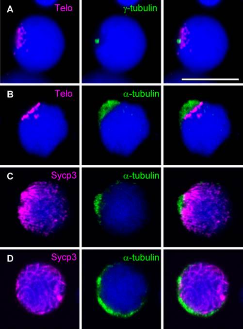
Fig. 6
Localizations of α-tubulin and ?-tubulin in spermatocytes. A: FISH using a telomere probe in combination with immunocytochemical labeling of ?-tubulin. B: FISH using a telomere probe in combination with immunocytochemical labeling of α-tubulin. C,D: Double immunocytochemical labeling of α-tubulin and Sycp3 at the early zygotene (C) and zygotene (D) stages. α-Tubulin staining expands during the zygotene stage. Left panels show telomere or Sycp3 staining (magenta), middle panels show ?-tubulin or α-tubulin staining (green), and right panels show merged images of the left and middle panels. Nuclei were stained with TO-PRO-3 (blue). Scale bar = 10 �m.