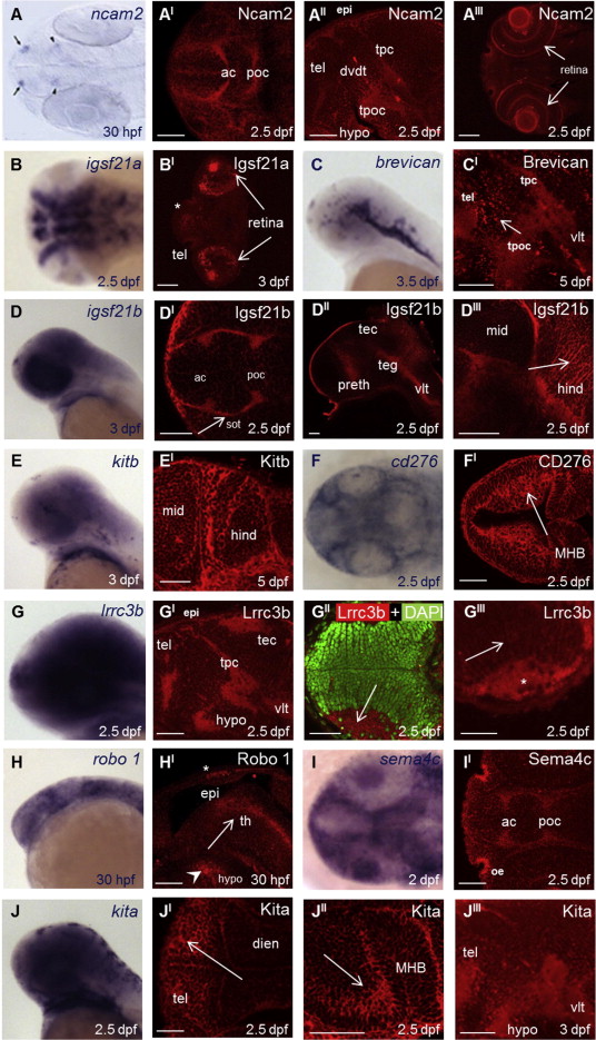
Fig. 2
Immunohistochemistry of the embryonic zebrafish brain with monoclonal antibodies to neural receptors and secreted proteins. The embryo stage and transcript/protein are indicated within each subpanel and staining patterns described within the main text. Transcript localisation is shown by wholemount in situ hybridisation as brightfield images; images of antibody staining are taken by confocal microscopy (red); nuclei are stained with DAPI in GII (green). Note that some antibody staining, for example Igsf21a in the ventral diencephalon (BI), is outside of the optical plane. Orientation of the embryos is always anterior to the left with ventral views in A, AI, DI, II and JI; lateral in AII, C, CI, D, DII, DIII, E, EI, G, GI, H, HI, J and JIII; and dorsal in AIII,B, BI, F, FI, GII, GIII, I and JII. Scale bars represent 100 Ám in CI and EI, for all other images 50 Ám. Abbreviations used in all figures are: ac = anterior commissure, dvdt = dorso?ventral diencephalic tract, epi = epithalamus, hind = hindbrain, hypo = hypothalamus, MHB = mid-hindbrain boundary, mid = midbrain, ob: olfactory bulb, oe = olfactory epithelium, poc = postoptic commissure, sot = supraoptic tract, tec = tectum, teg = tegmentum, tel = telencephalon, th = thalamus, tpc = tract of the posterior commissure, tpoc = tract of the postoptic commissure, vlt = ventral longitudinal tract, vnc = ventral nerve cord, hpf = hours post-fertilisation, dpf = days post-fertilisation.

