Fig. 5
Apoptosis, DNA damage signaling and fibrosis in zebrafish.
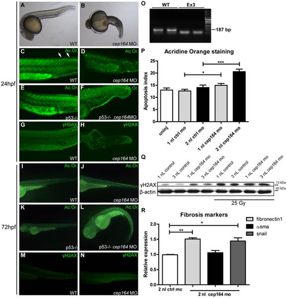
Zebrafish cep164 knockdown leads to developmental abnormalities associated with apoptosis and DNA damage. A cep164 morpholino targeting the exon 3 splice donor reduces wildtype mRNA (lanes WT) and results in a smaller cep164 RT-PCR product indicating an internal deletion (lanes Ex3) (O) was injected into 1?2 cell embryos. Compared to wild-type embryos at 24 hpf (A), morphant embryos (B) were stunted and displayed shorten body axis, body axis curvature, and edema. No cysts or other specific phenotypes were observed in the pronephros of morphant fish. Acridine orange staining revealed a low level of apoptosis in control injected WT (C) and p53 -/- embryos (E) restricted primarily to the posterior neural tube (arrows in C). cep164 knockdown induced widespread apoptosis (D) in the trunk and tail which was not affected by p53-deficiency as indicated by the cep164 knockdown-induced apoptosis in p53-/- mutants (n = 30) (F). Staining with ?H2AX antibody revealed enhanced DNA damage signaling in cep164-deficient embryos (n = 10) (H) but not controls (G). Acridine orange staining (Ac. Or.) also revealed a low level of apoptosis in 72 hours post fertilization (hpf) control injected WT (I) and p53-/- embryos (K). cep164 knockdown induced widespread apoptosis (J) which was not affected by p53-deficiency as indicated by the cep164 knockdown-induced apoptosis in p53-/- mutants (n = 45) (L). Quantified acridine orange staining (Ac. Or.) reveals significantly increased apoptosis in WT zebrafish after cep164 knockdown (P). Student′s t-test were used to calculate p-values (*<0.05, ***<0.001) (n = 70 in 4 experiments, error bars represent SEM). Staining with ?H2AX antibody revealed enhanced DNA damage signaling in cep164-deficient embryos (N) but not control injected WT embryos (n = 10) (M). Western blot of ?H2AX protein levels from lysates of 15 pooled embryos, two hours after irradiation with 25 Gy, show increased DNA damage signaling (Q). RT-QPCR reveals significant induction of snail and fibronectin1 in cep164 MO injected embryos at 32 hpf (R). mRNA expression from 12 pooled embryos is normalized to 2 nL control MO injected zebrafish. Student′s t-test was used to calculate p-values (*<0.05, **<0.01).

