
|
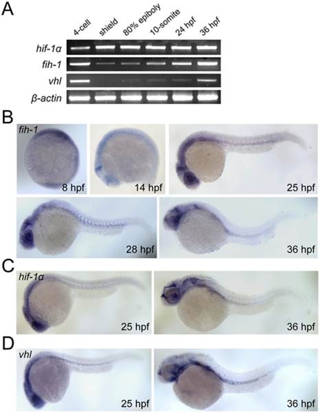
Fig. 2
Expression pattern of fih-1, vhl, and hif-1α.
(A) RT-PCR analysis showing expression of fih-1, hif-1a, vhl, and β-actin during development. (B) Whole-mount in situ hybridization of fih-1 during development. As early as 8 hpf, fih-1 expression can be detected. At 14 hpf, fih-1 is strongly expressed at the midbrain-hindbrain boundary and eye. At 25 hpf, expression of fih-1 is expanded to the optic vesicle and ventral mesoderm. The expression of fih-1 in midbrain-hindbrain boundary, eye, optic vesicle and ventral mesoderm is maintained at later stages. (C) Expression of hif1α, a known target of Fih-1, at 25 hpf and 36 hpf. (D) Expression of vhl, which is known to interact and synergize with Fih-1, at the same developmental stage. Both hif1α and vhl express within the similar anatomical region as fih-1.