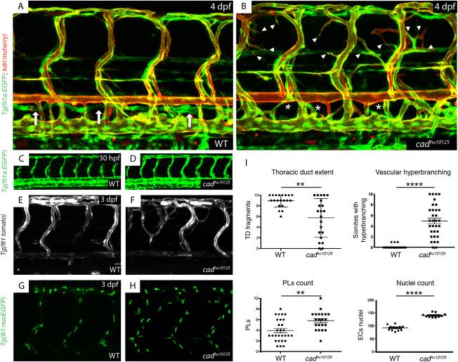
Fig. 1 hu10125 mutants present with lymphatic vascular deficiency and arterial hyperbranching. A,B: The vasculature Tg(fli1a:EGFP; flt1:tomato) in wild-type siblings (A) and hu10125 embryos (B) at 4 dpf (white arrows indicate a thoracic duct, asterisk an absence of thoracic duct, and arrowheads indicate hyperbranching). C,D: The vasculature Tg(fli1a:EGFP; flt1:tomato) in wild-type siblings (n = 53) (C) and hu10125 (n = 12) embryos (D) at 30 hpf. E,F: The arteries Tg(flt1:tomato) in wild-type siblings (C) and hu10125 (D) embryos at 3 dpf. G,H: Endothelial nuclei Tg(fli1:nucEGFP) in wild-type siblings (G) and hu10125 (H) embryos at 3 dpf. I: Quantification of: thoracic duct extent (number of thoracic duct fragments visible across 10 segments in the trunk) in WT (n = 20) and hu10125 (n = 21) (4 dpf); vascular hyperbranching (number of bilateral paired ISVs presenting with extra branches across 10 segments in the trunk) in WT (n = 32) and hu10125 (n = 32) (4 dpf); parachordal lymphangioblasts (number of PLs visible at the level of the horizontal myoseptum, across 10 segments in the trunk) in WT (n = 28) and hu10125 (n = 21) (56 hpf); and nuclei count (in ISVs, scored dorsal to the horizontal myoseptum across 10 segments) in WT (n = 12) and hu10125 (n = 10) embryos (3 dpf).
Image
Figure Caption
Figure Data
Acknowledgments
This image is the copyrighted work of the attributed author or publisher, and
ZFIN has permission only to display this image to its users.
Additional permissions should be obtained from the applicable author or publisher of the image.
Full text @ Dev. Dyn.

