
Image
|
Figure Caption
Fig. 3
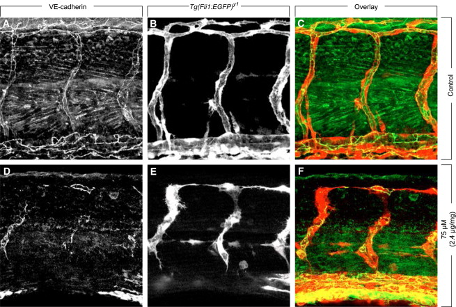
Inhibition of FGFRs Results in Reduced VE-Cadherin Junctions
Immunostaining of whole-mount zebrafish embryos for VE-cadherin (A and D, or red in C and F), showing intact and continuous EC-cell contacts in control-treated Tg(fli1:EGFP)y1 zebrafish embryos at 49 hpf (A?C), while SSR-treated zebrafish (exposure to 75 μM in the swimming water; tissue concentration 2.4 μg SSR/mg protein) (D?F) had a discontinuous VE-cadherin lining with reduced VE-cadherin-positive EC-cell juctions, explaining why ECs became disconnected from each other.
Figure Data
Acknowledgments
This image is the copyrighted work of the attributed author or publisher, and
ZFIN has permission only to display this image to its users.
Additional permissions should be obtained from the applicable author or publisher of the image.
Full text @ Chem. Biol.