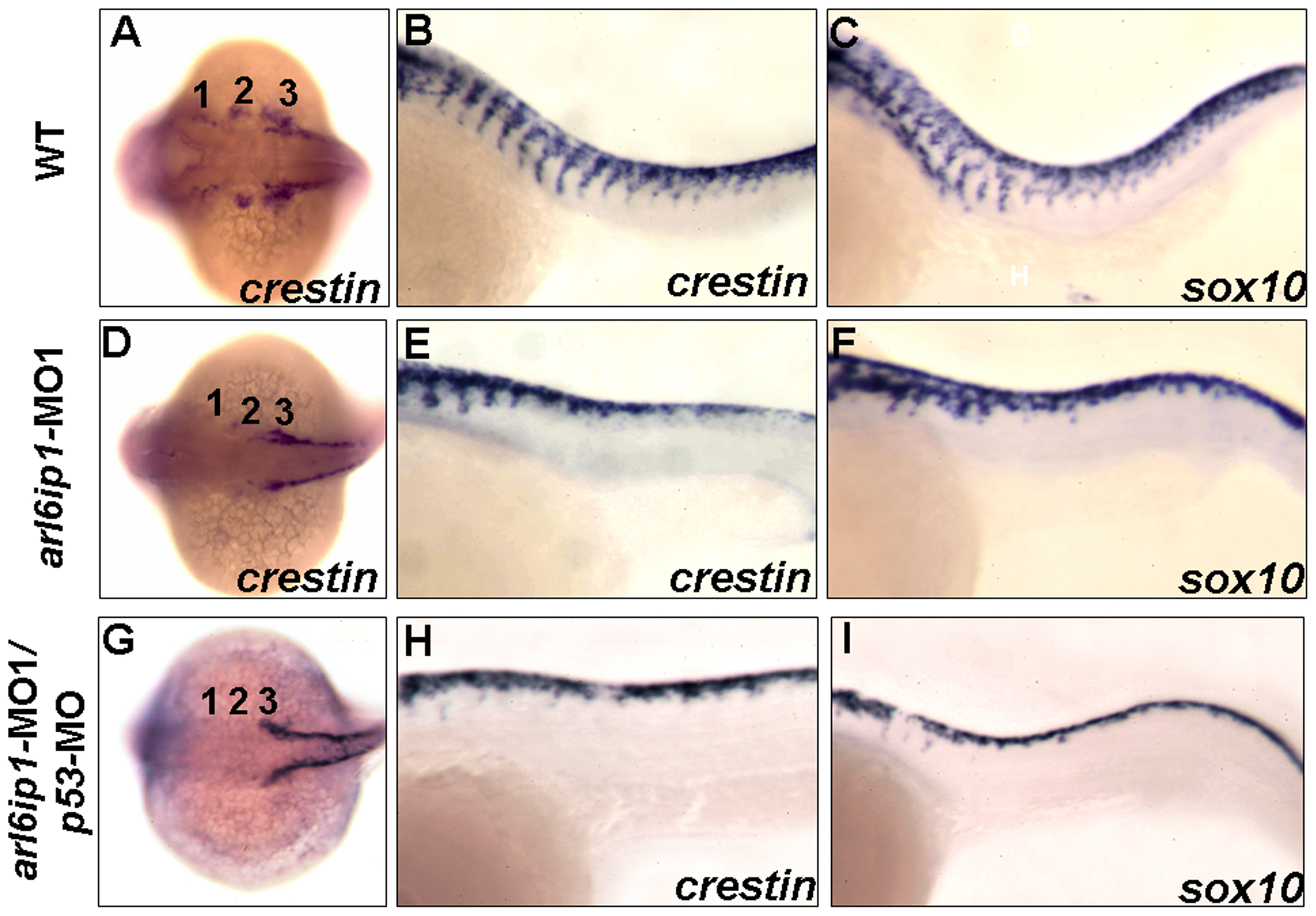
Fig. 6 Abnormal cell migration in arl6ip1-MO1-injected embryos.
Wild-type (A-C), arl6ip1-MO1-injected (D-F) and arl6ip1-MO1/p53-MO-injected (G-I) embryos were shown under dorsal views at 20-hpf (A, D, G) and under lateral views at 26-hpf (B, C, E, F, H, I), anterior to the left. Crestin expression revealed the migration of the three cranial neural crest streams in the wild-type embryos at 20 hpf (A). In arl6ip1 and arl6ip1/p53 knockdown embryos, crestin expression in the cranial crest was reduced in the 3rd neural crest stream and almost absent in the 1st and 2nd stream (D, G). At 26 hpf, the trunk migratory neural crest cells in wild-type embryos, as labeled by crestin and sox10, gradually migrated to ventral (B, C). However, the sox10- and crestin-expressing cells in arl6ip1 knockdown and arl6ip1-MO1/p53-MO-injected embryos failed to migrate ventrally (E, F, H, I).

