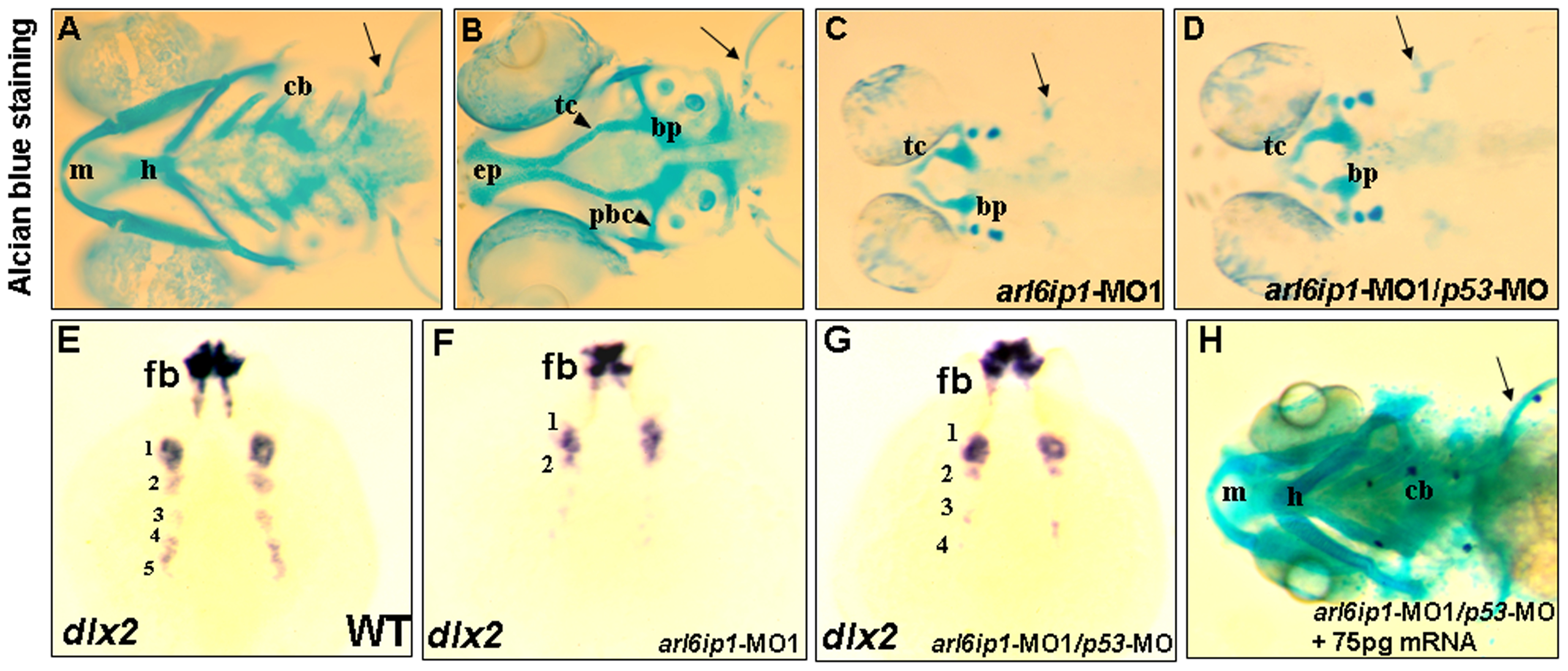
Fig. 2 Craniofacial cartilage defects in the arl6ip1 morphants.
Ventral views (A, C, D, H) or dorsal view (B) of 4-dpf embryos stained with Alcian blue to reveal craniofacial cartilage. Either pectoral fin (pf) or cartilage derived from mandibular (1st arch, m), hyoid (2nd arch, h), and ceratobranchial (3rd-7th arches, cb) arches was observed in control embryos (A). Dorsal views of anterior neurocranial cartilage in wild-type embryos showed paired trabeculae (tc) and an ethmoid plate (ep) (B). However, craniofacial cartilage and pectoral fin were absent in the arl6ip1-deficient (C) and arl6ip1-MO1/p53-MO-injected embryos (D). In the anterior neurocranial cartilage, trabeculae were severely reduced in both of these embryos, and the ethmoid plate was absent (C, D). dlx2 was expressed in migratory neural crest precursors of pharyngeal arch, as shown in dorsal views at 36 hpf in wild-type (E). Both arl6ip1-MO1-injected (F) and arl6ip1-MO1/p53-MO-injected embryos (G) had reduced dlx2 expression, and the reduction of dlx2 was prominent in posterior arches at 24 hpf. In addition, dlx2 was expressed normally in the forebrain (E, F, G). The severe chondrocranium and pectoral fin defects in arl6ip1-MO1/p53-MO-injected embryos were rescued by injection of 75 pg wobble arl6ip1 mRNA (H). bp, basal plate; pbc, posterior basic capsular commiss. Arrows, pectoral fin.

