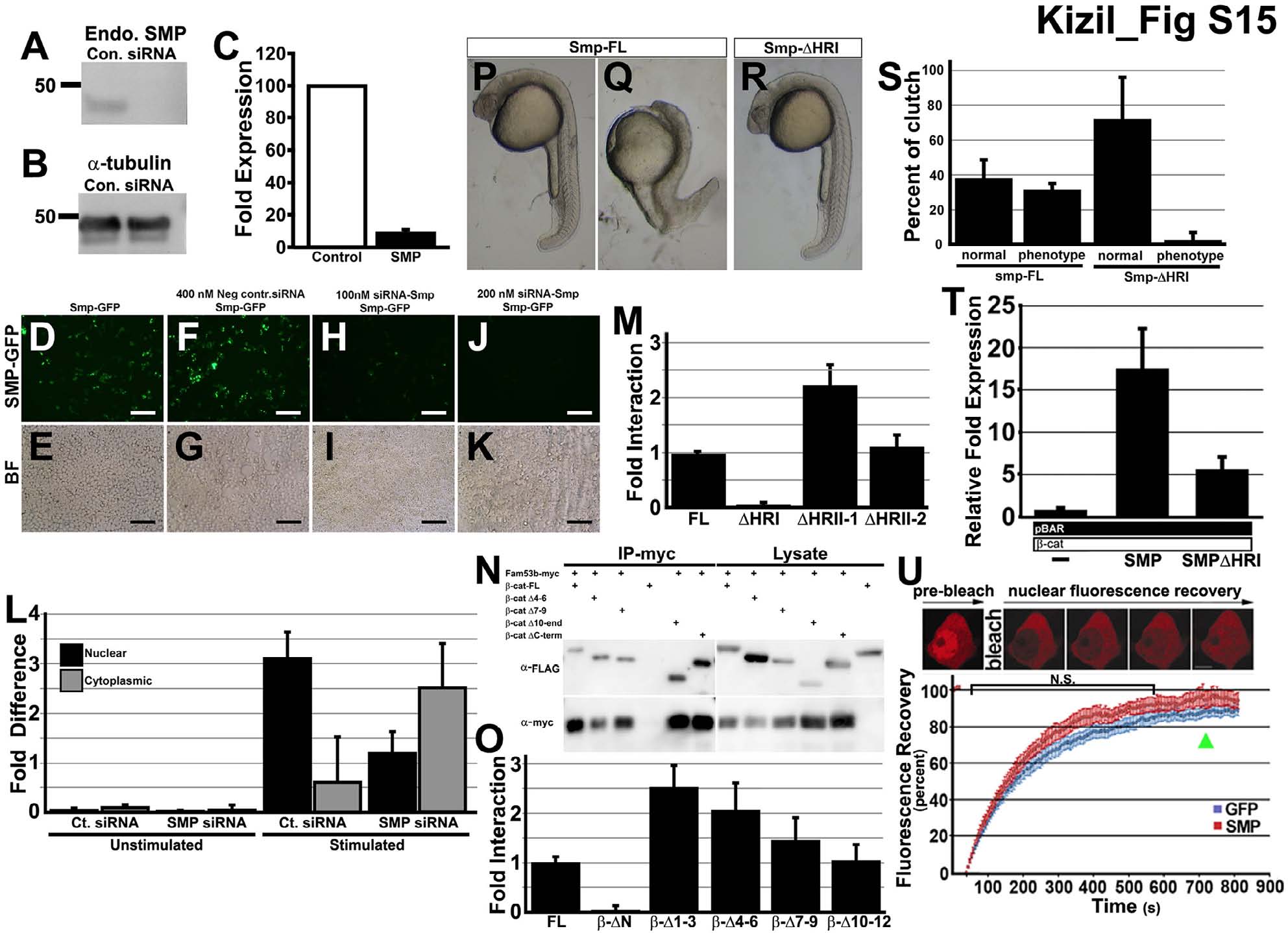
Fig. S15 Reduction of SMP protein by transfection of Smp siRNA, Related to Figure 4. (A) Western blot of immnoprecipitation pull down of β-catenin with different mutants of the Smp protein. (B) Western blot of antibody to the endogenous SMP protein in control siRNA- and SMP siRNA-transfected HEK293T cells. (C) Same blot samples probed with α-tubulin antibody. (D) Measurement of fold expression detected from Western blot of siRNA knockdown experiment. (E-L) Expression of GFP fused to the C-terminal of Smp. (E) Smp-GFP alone and (F) Bright field image of (E). (G) Smp-GFP in cells transfected with control siRNA. (H) bright field of (G). (I) SMP-GFP fluorescence in the cells transfected with 100 nM siRNA to Smp. (K) SMP-GFP in cells transfected with 200 nM of siRNA to Smp. (M) Lucifierase reporter assay for control β-catenin, Smp-FL and Smp delta homology region (I). (N) Fluorescence recovery curves after photobleaching mCherry-β-catenin in nucleus of cell transfected either with GFP or Smp. Scale bars equal 100 μm.
Image
Figure Caption
Acknowledgments
This image is the copyrighted work of the attributed author or publisher, and
ZFIN has permission only to display this image to its users.
Additional permissions should be obtained from the applicable author or publisher of the image.
Full text @ Development

