
|
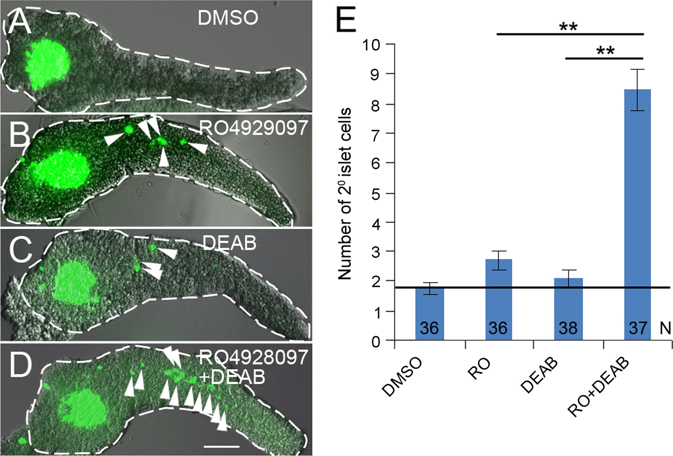
Fig. 1
Notch and RA signaling regulate the induction of 2� islet cells in a manner consistent with synergy. (A?D) Confocal z-stack projections of larval Tg(neuroD:GFP)nl1 pancreata dissected at 5 dpf. Larvae were treated with (A) DMSO, (B) RO4929097 (RO) 0.625 μM, (C) DEAB 25 μM and (D) DEAB 25 μM+RO 0.625 μM, from 3?5 dpf. 2� Islet cells (green) in the tail region of pancreata are indicated by arrowheads. Pancreata are outlined in white dashed lines. Scale Bar, 100 μm. (E) Average number of 2� islet cells per 5 dpf larval pancreas. N=number of larval pancreata quantified. Error Bar represents standard error around the mean (SE). Significance, **p<0.001.
Reprinted from Developmental Biology, 394(1), Huang, W., Wang, G., Delaspre, F., Vitery, M.D., Beer, R.L., Parsons, M.J., Retinoic acid plays an evolutionarily conserved and biphasic role in pancreas development, 83-93, Copyright (2014) with permission from Elsevier. Full text @ Dev. Biol.