
|
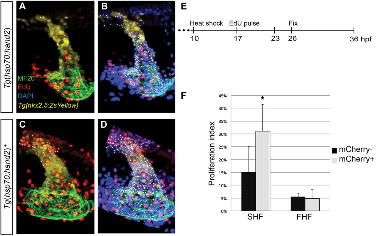
Fig. 6
Overexpression of hand2 increases proliferation of SHF-derived progenitor cells. (A-D) EdU incorporation in (A,B) nontransgenic and (C,D) Tg(hsp70:hand2) embryos at 26hpf, following heat shock at 10hpf and EdU pulse at 17hpf; partial reconstructions of confocal z-stacks with ventricle upwards. (A,C) White dots indicate EdU-positive (red) cells that are MF20 positive (green) and/or expressing Tg(nkx2.5:ZsYellow) (yellow). (B,D) White dots indicate all nuclei (DAPI, blue) of cells that are MF20 positive and/or expressing Tg(nkx2.5:ZsYellow). (E) Timeline of experimental design. (F) Proliferation indices, as in Fig. 5H, for two populations of cells: SHF-derived cells, defined as Tg(nkx2.5:ZsYellow)-expressing cells with very low or no MF20 staining; and FHF-derived cells, defined as cells with clearly detectable MF20 staining. Proliferation index was calculated for each population independently by dividing the number of EdU-positive cells by the total number of cells in the population. A significant increase in proliferation index was evident in the SHF-derived cells in hand2-overexpressing embryos (n=10 or 11; *P=0.003), but not in the FHF-derived cells (n=10 or 11; P=0.528).