
|
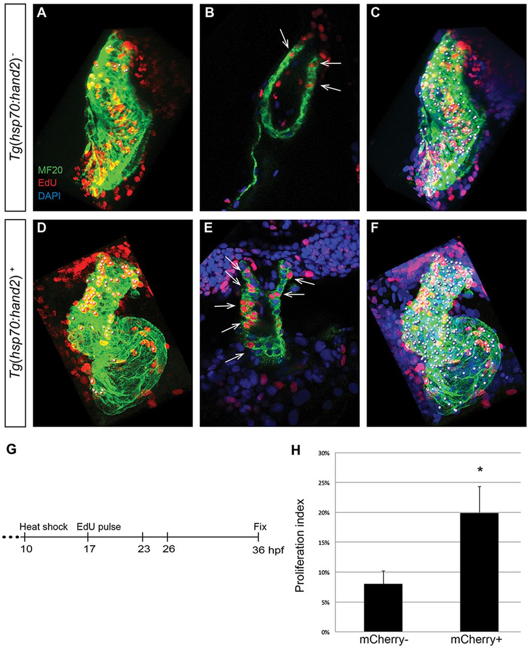
Fig. 5
Increased proliferation contributes to cardiac expansion in hand2-overexpressing embryos. (A-F) EdU incorporation in hearts of (A-C) nontransgenic and (D-F) Tg(hsp70:hand2) embryos at 36hpf, following heat shock at 10hpf and EdU pulse at 17hpf. Partial reconstructions of confocal z-stacks with ventricle upwards (A,C,D,F) and representative single slices (B,E). (A,D) White dots indicate EdU-positive (red) cells that are also MF20-positive (green) differentiated cardiomyocytes. (B,E) Arrows indicate EdU-positive cells that are also MF20 positive; DAPI (blue) marks all nuclei. (C,F) White dots indicate all myocardial nuclei. Qualitative assessment suggests increased numbers of EdU-positive cardiomyocytes in hand2-overexpressing hearts (D,E), particularly in the distal region of the ventricle and the outflow tract. (G) Timeline of experimental design. (H) Proliferation indices in nontransgenic (mCherry-negative) and Tg(hsp70:hand2) (mCherry-positive) embryos; error bars indicate s.d. Proliferation index was calculated by dividing the number of EdU-positive cardiomyocytes by the total number of cardiomyocytes. A significant increase in proliferation index was evident in hand2-overexpressing embryos (n=8 or 9; *P<0.001).