Fig. 3
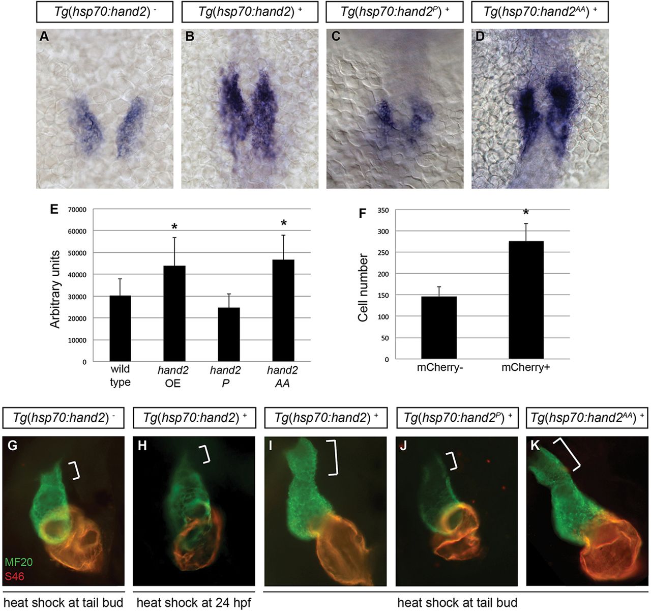
Cardiac expansion induced by hand2 overexpression requires phosphorylation-independent dimerization of Hand2. (A-D) In situ hybridization depicts cmlc2 expression at 18 somites in (A) nontransgenic embryos, (B) Tg(hsp70:hand2) embryos, (C) Tg(hsp70:hand2P) embryos and (D) Tg(hsp70:hand2AA) embryos, heat-shocked at 10hpf; dorsal views, anterior upwards. (E) Average area of cmlc2 expression, as in Fig. 1E, in nontransgenic and transgenic embryos, following heat shock at 10hpf. Asterisks indicate significant differences from nontransgenic embryos. Overexpression of hand2 or hand2 AA increases the area of cmlc2 expression (n=15-32; *P<0.001), whereas hand2 P does not (n=13; P=0.05). (F) Average number of cardiomyocytes at 36hpf, as in Fig. 1F, in nontransgenic embryos and in Tg(hsp70:hand2) embryos, following heat shock at 10hpf. Asterisk indicates a significant difference from nontransgenic embryos (n=16-20; *P<0.001). (G-K) Immunofluorescence at 36hpf for MF20 (green, visible in the ventricle) and S46 (red, visible in the atrium) shows cardiac morphology. Frontal views; brackets mark the outflow tract. Hearts of Tg(hsp70:hand2) embryos heat-shocked at 24hpf (H) resemble nontransgenic hearts heat-shocked at 10hpf (G). Tg(hsp70:hand2) embryos heat-shocked at 10hpf (I) show an overall increase in cardiac size and an enlarged outflow tract. Similar morphology is seen after heat shock at 10hpf in Tg(hsp70:hand2AA) embryos (K), but not in Tg(hsp70:hand2P) embryos (J).

