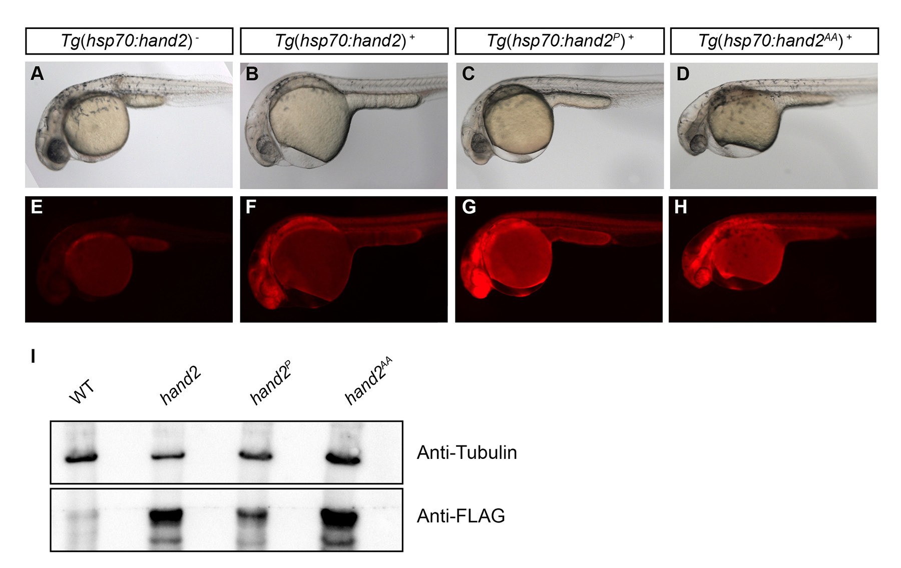
Fig. S3
Levels of protein production in different transgenic lines. (A-H) Lateral views of live embryos at 30 hpf depict bright field images of representative embryos from different transgenic lines (A-D), along with corresponding mCherry fluorescence (E-H). (E) No mCherry fluorescence is induced in heat-shocked nontransgenic embryos. (F-H) mCherry fluorescence is readily detectable in representative embryos carrying the transgenes (F) Tg(hsp70:FLAG-hand2-2A-mCherry) (Tg(hsp70:hand2)), (G) Tg(hsp70:FLAG-hand2P-2A-mCherry) (Tg(hsp70:hand2P)), and (H) Tg(hsp70:FLAG-hand2AA-2A-mCherry) (Tg(hsp70:hand2AA)). (I) Western blot analysis compares levels of FLAG-tagged Hand2 protein in different transgenic lines. Embryos were deyolked and lysates were prepared as previously described (Link et al., 2006), and blots were probed with either a monoclonal anti-FLAG M2 antibody (F1804, Sigma, 1:2000) or a monoclonal anti-α-Tubulin antibody (T6728, Sigma, 1:10,000), followed by a rabbit anti-mouse IgG HRP-conjugated secondary antibody (ab97046, Abcam, 1:10,000). Proteins were visualized using SuperSignal West Femto Chemiluminescent Substrate (Thermo Scientific). Each lane contains lysate from 15 embryos at 36 hpf (2 hours following heat shock), and the lanes compare protein levels in nontransgenic embryos (WT), Tg(hsp70:hand2) embryos (hand2), Tg(hsp70:hand2P) embryos (hand2P), and Tg(hsp70:hand2AA) embryos (hand2AA). All three transgenic lines contain a ~23 kD FLAG-Hand2 protein, with comparable levels in Tg(hsp70:hand2) and Tg(hsp70:hand2AA) embryos and lower levels in Tg(hsp70:hand2P) embryos.

