
|
Fig. 3
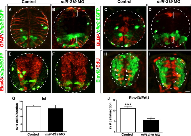
miR-219 Is Required for Differentiation of Glia and Late-Born Neurons
All images show representative transverse sections through trunk spinal cord with dorsal up.
(A and B) Whereas numerous GFAP+ radial glia occupy the spinal cord of a 3 dpf control larva, a miR-219 MO-injected larva has few radial glia except for the most dorsal and ventral regions of spinal cord (brackets). Asterisks mark oligodendrocyte lineage cells.
(C and D) Images showing a deficit of BLBP+ radial glia in a miR-219 MO-injected larva compared to control.
(E and F) miR-219 MO-injected larvae appear to have a normal number and distribution of neurons, marked by Elavl3 expression, but fewer oligodendrocyte lineage cells (asterisks) than control larvae.
(G) Graph showing number of Isl+ motor neurons in control and miR-219 MO-injected larvae. Data are presented as mean � SEM (n = 10 sections obtained from 15 larvae per group, with two replicates). p > 0.05, unpaired t test.
(H and I) Confocal images of embryos pulsed with EdU at 1 dpf and fixed at 2 dpf. Numerous Edu+ cells are also Elavl3+ (arrowheads) in control larvae (H), whereas most EdU label persists within cells lining the spinal cord lumen, and fewer neurons are labeled by EdU in miR-219 MO-injected larva (I).
(J) Graph showing number of Elav3+ EdU+ neurons in control and miR-219 MO-injected larvae. Data represent mean � SEM (n = 10 sections obtained from five larvae per group). p < 0.0001, unpaired t test. Scale bar equals 10 μm.
Reprinted from Developmental Cell, 27(4), Hudish, L.I., Blasky, A.J., and Appel, B., miR-219 Regulates Neural Precursor Differentiation by Direct Inhibition of Apical Par Polarity Proteins, 387-398, Copyright (2013) with permission from Elsevier. Full text @ Dev. Cell