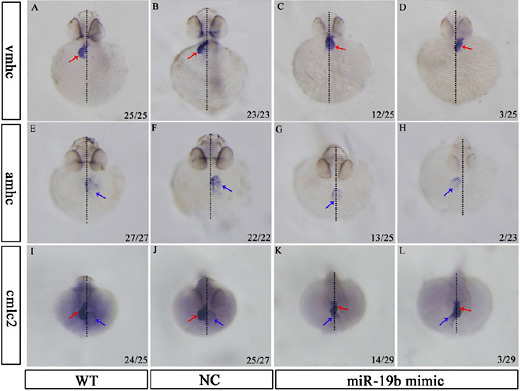
Fig. 7
Overexpression of miR-19b alters the expression of cardiac chamber marker genes. Representative images (8 times magnification) of 48 hpf embryos showing the expression of vmhc (A, B, C and D), amhc (E, F, G and H) and cmlc2 (I, J, K and L) by in situ hybridization are shown. After the heart tube has formed, leftward movement of the heart (jogging) is followed by rightward bending of the ventricle (D-looping) in WT embryos (A, E and I) and NC-injected embryos (B, F and J). In contrast, miR-19b mimic-injected embryos exhibited multiple LR defects, including reversed looping (L-looping; D, H and L) and no looping (C, G and K). In the bottom right-hand corner, the number of embryos examined with the representative expression/total number of embryo are presented. Black dashed lines: the midline of the body axis; red arrow: ventricle; blue arrow: atrium.

