
|
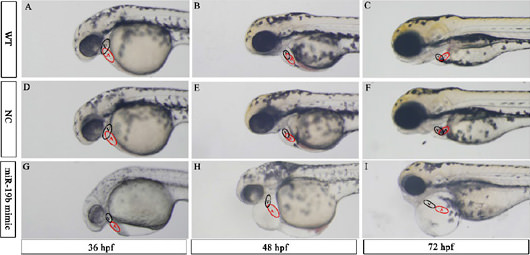
Fig. 5
Overexpression of miR-19b leads to embryonic defects in zebrafish cardiac morphology. Representative images (6.3 times magnification) of the lateral view of WT (A, B and C), NC (D, E and F), and mir-19b mimic injected embryos (G, H, and I) are shown. In WT and NC embryos, the ventricle and atrium largely overlap with each other; however, the ventricles of mir-19b mimic-injected embryos were positioned anterior to the atrium with little overlap. Instead of the normal looped and S-shaped hearts observed in the control group, the heart chambers of miR-19b mimic-injected embryos were string-like and elongated. V: ventricle; A: atrium.