Fig. 2
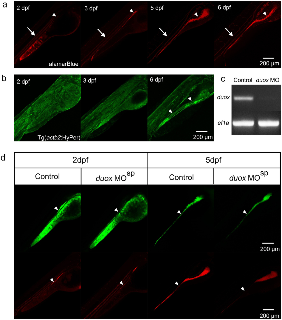
(a) The staining patterns of almarBlue reveal the gut lumen (white arrowheads) and circulating blood cells (white arrows) at 2?6 dpf in the lateral view. (b) Green signals are universally detected in Tg(actb2:HyPer)pku326 before 3 dpf, and the signals enhance in the intestinal epithelial cells at 6 dpf (white arrows). (c) RT-PCR reveal the efficient block of duox transcript splicing via MO mediated genetic knockdown. (d) The signals of the ROS/redox probes reduce, but not exclusively disappear, in the intestinal tract after duox is genetic knockdown by MO. White arrowheads indicate the signals in the intestinal tract.

