Image
Figure Caption
Fig. S2
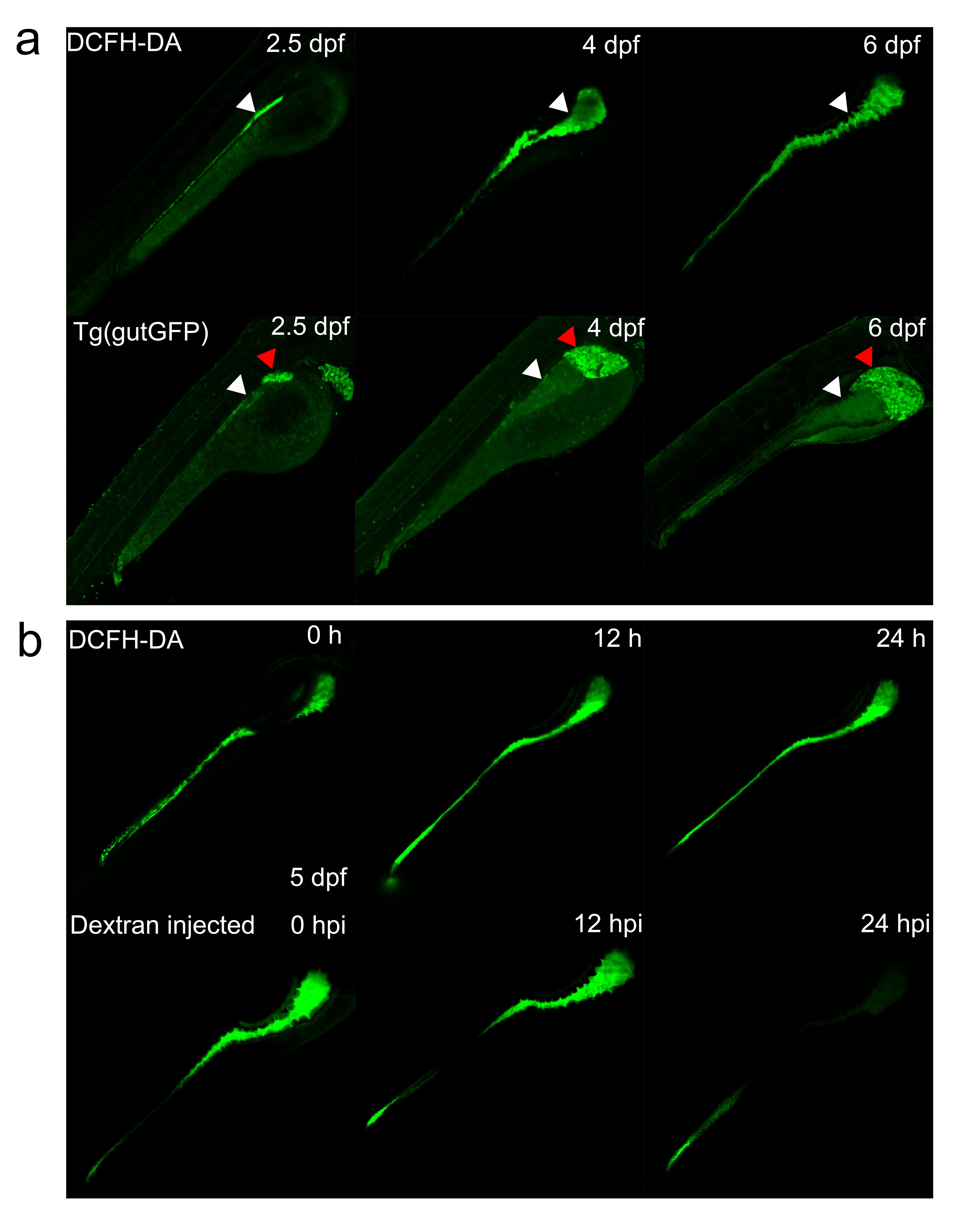
Comparison of different intestinal detection assays.
(a) The reflection of intestine by DCFH-DA and Tg(gutGFP) at different stages. White arrowheads indicate the intestine tract, whereas red arrowheads represent the liver in Tg(gutGFP). (b) The intensity of signals decreases significantly with time after injection of Dextran into the intestine at 5 dpf.
Acknowledgments
This image is the copyrighted work of the attributed author or publisher, and
ZFIN has permission only to display this image to its users.
Additional permissions should be obtained from the applicable author or publisher of the image.
Full text @ Sci. Rep.

