Fig. 7
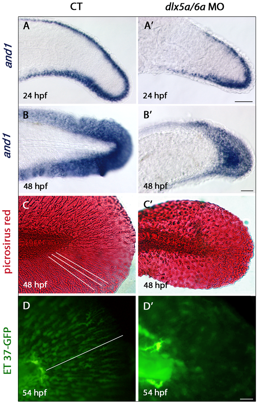
Defects in actinotrichia formation in the median fin fold of dlx5a/6a morphants.
Lateral views of the developing median fin fold (MFF) of control (A?D) and dlx5a/6a morphant (A2?D2) embryos. Whole mount in situ hybridization for and1 at 24 (A, A2) and 48 hpf (B, B2). In control embryos, and1 is expressed in epithelial cells of the fin fold at 24 hpf and in all MFF cells at 48 hpf (A?B). In dlx5a/6a morphants, and1 expression is detected in the MFF but transcripts show a less extended antero-posterior expression domain at 24 hpf (A2). At 48 hpf, morphants display a thinner and1 expression domain in the ventral and dorsal part of the MFF (B2). The picrosirius red histological staining in 48 hpf control embryos reveals the MFF cellular organisation with the presence of actinotrichia (C, the white lines indicate their orientation and length). In the morphants, the MFF is smaller and granular associated with absence of actinotrichia (C2). The ET 37-GFP enhancer-trap line reveals the MFF mesenchymal cells displaying filopodia which migrate along the actinotrichia at 54 hpf (D, the white line indicates the direction of migration). The dlx5a/6a knock down in ET 37-GFP embryos leads to impaired MFF mesenchymal migration, the cells are disorganized and do not show filopodia. Scale bars in A?B 50 μm, C 20 μm, D 10 μm.

