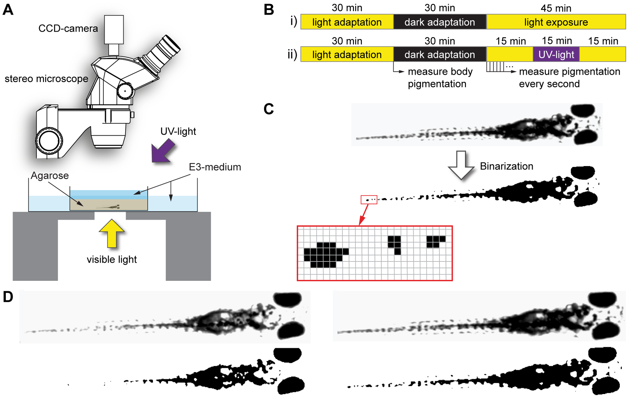
Fig. 1
Experimental procedure.
A) Schematic drawing of the experimental setup. Zebrafish larvae are embedded in agarose and placed under a dissecting microscope. Illumination with visible light is provided from below, optional near-ultraviolet light from obliquely above. B) After an initial light adaptation period lasting 30 minutes, the extent of body pigmentation is measured and serves as a reference for all following measurements. The larvae are then dark adapted for another 30 minutes, after which they are exposed to visible light for 45 minutes (i). During light exposure, the extent of pigmentation is measured every second. Optional UV light is provided during a 15 minutes interval in the middle of the light exposure phase (ii). C) To quantify the extent of body pigmentation, images of the larva are binarized using a fixed threshold. The number of pixels above this threshold is summed and divided by the number of pixels above threshold after 30 minutes light adaptation, resulting in a ?melanin index?. D) Example original (top row) and binarized (bottom row) images of the same 5 day old zebrafish larva with minimal melanosome dispersion after dark adaptation (left, melanin index = 1.14) and maximal melanosome dispersion reached after 15 minutes of UV exposure (right, melanin index = 1.98).

