
|
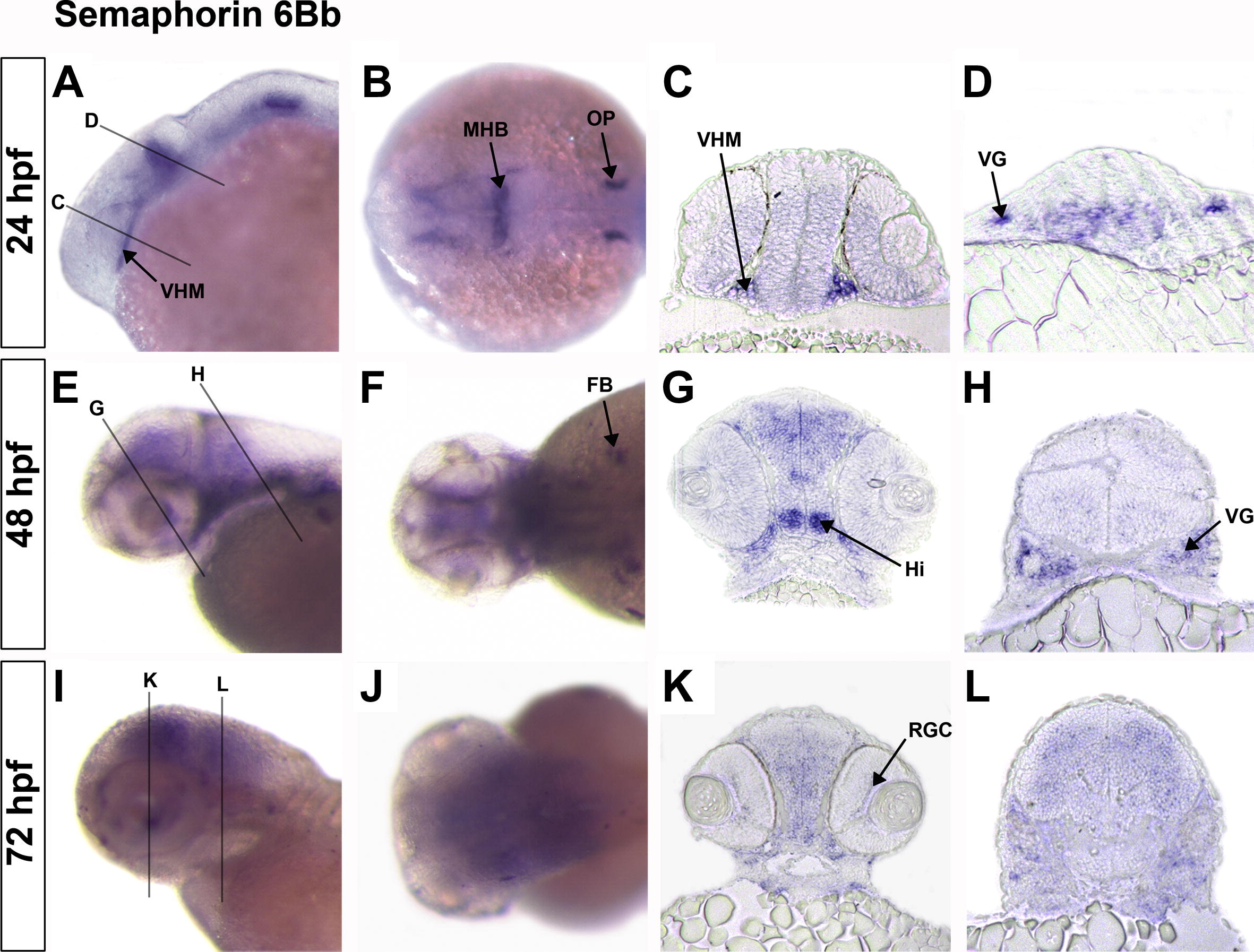
Fig. 4 In situ hybridization of sema6Bb gene expression at 24 hpf (A?D), 48 hpf (E?H) and 72 hpf (I?L). Lateral views (A, E, and I) with anterior to the left, dorsal at the top, dorsal views (B, F, and J) with anterior to the left, rostral transverse brain sections (C, G, and K) and caudal transverse brain sections (D, H, and L) with dorsal at the top. Parallel lines indicate where sections were imaged. Arrows indicate expression in the otic placode (OP), midbrain?hindbrain boundary (MHB), ventral head mesenchyme (VHM), vagal ganglion (VG), fin bud (FB), intermediate hypothalamus (Hi) and retinal ganglion cells (RGC).
Reprinted from Gene expression patterns : GEP, 12(3-4), Ebert, A.M., Lamont, R.E., Childs, S.J., and McFarlane, S., Neuronal expression of class 6 semaphorins in zebrafish, 117-122, Copyright (2012) with permission from Elsevier. Full text @ Gene Expr. Patterns