
|
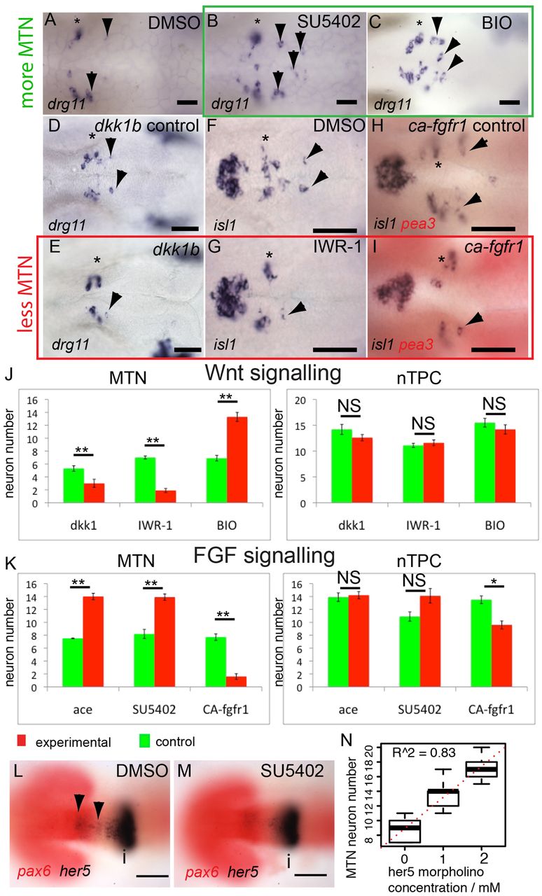
Fig. 2
FGF, Wnt and Her5 dictate the number of MTN neurons that form in the midbrain. In situ hybridisation with probes for drg11 (A-E) and isl1 (F-I) reveals increased numbers of MTN neurons in zebrafish embryos exposed to 40 μM SU5402 (B) or 4 μM BIO (C) from 14 hpf (green box), relative to DMSO treatment (A). Overexpression of Dkk1b in Tg[hsp70l:dkk1b-egfp] embryos (D,E), treatment with 40 μM IWR-1 (F,G) or overexpression of a constitutively active Fgfr1 in Tg[hsp70:ca-fgfr1] embryos (H,I) also results in fewer MTN neurons (red box). Arrowheads indicate MTN neurons; asterisks indicate nTPC neurons. Quantification of neurons in animals with altered FGF (K) or Wnt (J) activity in the midbrain reveals a highly significant increase in MTN (P<0.01) but not nTPC (P>0.01) neuron number at 24 hpf for all conditions. ace, acerebellar. her5 expression in the dorsal midbrain (arrowheads) is lost by 20 hpf after application of 20 μM SU5402 at 14 hpf (M), but isthmus expression if unaffected relative to DMSO-treated controls (L). Plots of MTN and nTPC numbers in Her5 morphants reveals that loss of Her5 function causes a dose-dependent increase in MTN (R2=0.83) by 24 hpf (N). Error bars indicate s.e.m. **P<0.01; *P<0.05; NS, not significant; unpaired t-tests were used to compare between conditions; n=10 for each condition. i, isthmus. Scale bars: 100 μm in L,M; 20 μm in A-I.