
|
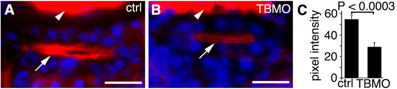
Fig. 6
Apical actin is reduced in the pronephric tubule epithelia of sept7b-knockdown larvae. (A,B) Staining with phalloidin shows prominent apical actin (arrows) in the pronephric tubule of larvae injected with the TBMO-MM control (ctrl; A). By contrast, the intensity of actin staining is reduced in larvae injected with the sept7b TBMO (TBMO, B). The arrowheads point to muscle with intense labeling for actin. DAPI (blue) stains the nuclei. (C) The fluorescence intensity of actin staining in the pronephric tubules is reduced in sept7b morphants. The same settings were used during imaging of control and morphant larvae, and the images were analyzed without processing. Scale bars: 10 μm.