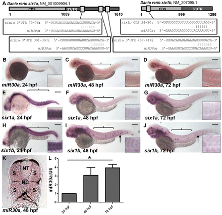
Fig. 1 miR30a displays a reciprocal expression pattern with six1a/b. (A) Schematic diagrams show predicted miR30a target binding sites in the 32 untranslated region (UTR) of six1a and the coding sequence (CDS) of six1b. (B?J) In situ hybridization reveals a lack of mature miR30a expression at 24 hpf (B), whereas mature miR30a localizes to the somites (indicated by brackets) at 48 and 72 hpf (C,D). This miR30a expression is reciprocal to expression of six1a (E?G) and six1b (H?J) transcripts observed in the developing somites at 24 hpf (E,H), which is lost at 48 hpf (F,I) and 72 hpf (G,J). Scale bars: 250Ám. Anterior is shown to the left; arrows, neuromasts. Insets show a higher magnification of somites above the yolk sac. (K) In situ hybridization for miR30a in 18-Ám transverse sections of embryos at 48 hpf reveals miR30a expression specifically in the somites (S), as compared to a lack of expression in the neural tube (NT). NC, notochord. (L) A quantification of miR30a expression performed by real-time PCR analysis shows an increase in miR30a expression from 24 to 72 hpf in tails from wild-type embryos. *P<0.027, ANOVA with Bonferroni′s post-hoc test.
Image
Figure Caption
Figure Data
Acknowledgments
This image is the copyrighted work of the attributed author or publisher, and
ZFIN has permission only to display this image to its users.
Additional permissions should be obtained from the applicable author or publisher of the image.
Full text @ J. Cell Sci.

