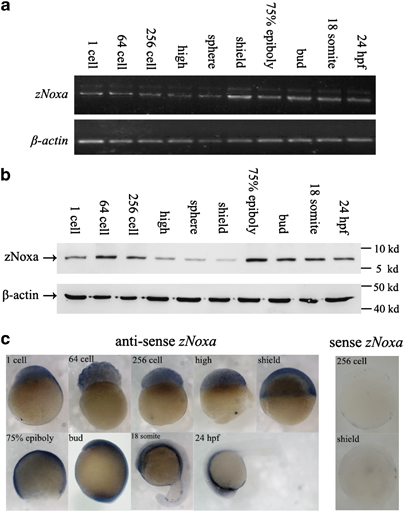
Fig. 1
Expression of zebrafish Noxa (zNoxa) during embryogenesis. (a) Transcription of zNoxa in embryonic development stages was detected by semi-quantitative RT-PCR and normalized to β-actin transcript. (b) The extracted proteins from different stage embryos were subjected to western blot detection by the mouse polyclonal antiboies against zNoxa-mature protein and β-actin, respectively. (c) Expression pattern of zNoxa was detected by in situ hybridization using specific antisense or sense probes. No staining was present in embryos hybridized with sense probe (256 cell and shield representative stages shown). Embryo in ?24 h.p.f.? panel was lateral view, with dorsal toward the top and anterior to the left. The embryos in other panels were lateral views with animal pole toward the top and dorsal to the right.

